Download

1 / 38
490 likes | 1.32k Views
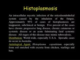
HISTOPLASMOSIS. HISTOPLASMOSIS. Darlings disease Causative fungus: Histoplasma capsulatum Disease of reticuloendothelial system Intracellular parasite Dimorphic fungus World wide in distribution but is most common in America. PATHOGENESIS

E N D
HISTOPLASMOSIS • Darlings disease • Causative fungus:Histoplasma capsulatum • Disease of reticuloendothelial system • Intracellular parasite • Dimorphic fungus • World wide in distribution but is most common in America
PATHOGENESIS • Source of infection :soil enriched with excreta of birds or bats • Route of infection :inhalation of spores
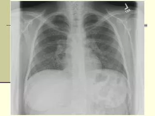
CLINICAL FEATURES • 90-95% are asymptomatic Acute pulmonary histoplasmosis : flu like symptoms malaise fever chills profuse sweating sore throat
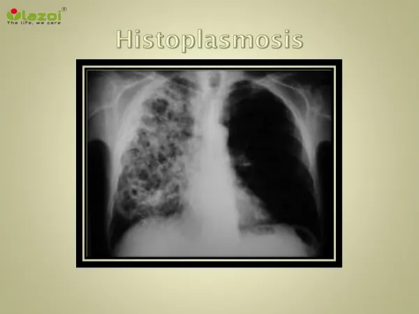
Continued…….. chest pain cough dyspnoea Chronic progressive pulmonary histoplasmosis: acute stage progresses leading to • haemoptysis • Apical and subapical cavities
Disseminated histoplasmosis: develops in minority of infected individuals • Involvement of RES leads to lymphadenopathy hepatosplenomegaly fever and anaemia • Cutaneous and mucocutaneous: granulomatous ulcerative lesions
LAB DIAGNOSIS SPECIMENS sputum bone marrow aspirate peripheral blood scrapings from ulcers biopsies of lymph nodes and other organs
DIRECT EXAMINATION • Smears of sputum or pus are stained with giemsa or wright stain • On microscopic examination H.capsulatum appears as small,oval yeast cell (2-4micron) Packed within the cytoplasm of macrophages or monocytes
CULTURE • SDA or brain heart infusion (BHI)agar with cycloheximide and chloramphenicol are inoculated • At 37c yeast phase is formed • At 25c appears as white cottony mycelial growth containing large(8-20microns)thick walled ,spherical spores with tubercles or finger like projections
SEROLOGICAL TESTS latex agglutination precipitation complement fixation They become positive 2 weeks after infection • Increase in titre of antibody indicates a progressive disease
HISTOPLASMIN SKIN TEST • Delayed hypersensitivity test • Similar to tuberculin test but antigen used is histoplasmin • Positive reaction indicates past or present infection,but does not differentiate active and passive infections
AFRICAN HISTOPLASMOSIS • Causative fungus:Histoplasma duboisii • Mainly confined with in the continent of Africa • Primarily involves skin and subcutaneous tissues • It is morphologically identical to H.capsulatum in its mycelial phase but differs in yeast phase
BLASTOMYCOSIS • Causative fungus:Blastomyces dermatitidis • Dimorphic fungus • Characterised by suppurative and granulomatous lesions particularly in lungs • Also effects skin,bone and genitourinary tract • North american blastomycosis
PATHOGENESIS • Route:inhalation • Source :soil containing spores CLINICAL FEATURES: PULMONARY BLASTOMYCOSIS: • Primary infection of lung may resemble TB or histoplasmosis • May be asymptomatic or may leads to focal consolidations,miliary lesions,abscess
CUTANEOUS BLASTOMYCOSIS • Primary lesion is papule secondary nodules ulcerative lesions DISSEMINATED • Mainly seen in immunocompromised individuals including AIDS
LAB DIAGNOSIS • Specimens sputum pus scrapings from skin lesions
DIRECT MICROSCOPY • 10%KOH mount thick walled yeast cells with a single broad based bud • H&E stain and PAS stains also show yeast cells in section
CULTURE • SDA or blood agar • At 25 c mycelial phase occurs slowly on incubation. filamentous with septate hyphae and many round or oval conidia • At 37 c yeast phase is seen-cells with thick,double contoured walls
Cultures should be incubated for atleast six weeks before discarding them as negative.
TREATMENT • Not recommended in asymptomatic cases • AMPHOTERICIN B • KETOCONAZOLE • ITRACONAZOLE