Download
1 / 13
130 likes | 411 Views
Case Study. Nitinol Implant That Fractured During Animal Trials. Background. Nitinol coronary device was observed to have fractured one month into a six-month animal trial Only fracture of over 30 devices in trial

E N D
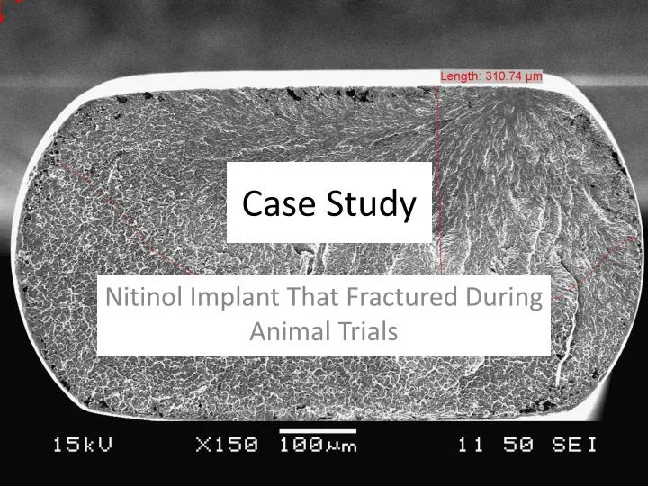
Case Study Nitinol Implant That Fractured During Animal Trials
Background • Nitinol coronary device was observed to have fractured one month into a six-month animal trial • Only fracture of over 30 devices in trial • Chemistry, transformation temperature, and mechanical property testing of the wire adjacent to the fracture indicated no issues • What happened??
Fracture Surface Analysis SEM examination to determine failure mode
Fracture Surface Analysis SEM examination of fracture surface mate
Fracture Surface Analysis Findings: • Fracture occurred due to fatigue crack initiation and growth • Fatigue crack initiated at an inclusion, propagated for approximately 314 microns before final fracture • No scratches or wear or other damage was observed near the fracture
Fracture Surface Analysis • Question: Did the inclusion “cause” this failure? • Answer: No, the inclusion presence, size, and location is typical in nitinol wire and acceptable per ASTM F 2063 for wrought nitinol mill products.
Analysis • So what caused the fracture? • Chemical, transformation, mechanical properties all checked out ok • Inclusions similar to the one that initiated the crack are expected (and would have been present in the other devices that passed testing) • What about loading?
Analysis • In vivo wire loads were reported to be primarily bending • Use linear-elastic fracture mechanics (LEFM) used to estimate loads, loading condition • Given crack length and fracture toughness, can calculate loads, bend radius at overload fracture • Conduct separate analyses for both bending and straight tension
Analysis • Nitinol fracture toughness values (non-embrittled) have been observed to range between 39 and 53 MPa(m)1/2 (He, et al. Smart Materials and Structures, Institute of Physics Publishing, 13, 2004, pp. N24-N28.) • LEFM analysis indicated: • 17 lb for final overload fracture – straight pull • Bend radius of 1.1 inches needed to induce final overload failure (pure bending > 70 ksi)
Analysis • LEFM results show straight pull unlikely • Bend radius reportedly within typical range for device in vivo • Therefore device fractured under apparently “typical” in vivo loading
Analysis What else could have caused the fracture? • Hydrogen? – Unlikely, would have likely affect transformation behavior • Residual stresses? – Bending to tight radii known to create tensile residual stresses in nitinol • Possibility of buckling wire during loading into catheter (plastic deformation/residual stress) • Fatigue testing of samples intentionally bent during simulated “push loading” showed significant life reduction • Loading procedure changed from “pushing” to “pulling”
Conclusions • Nitinol wire device failed by fatigue • In this case, inclusion at origin is not a “defect” • Chemistry, mechanical properties, transformation temperatures ok • LEFM indicated “typical” in vivo loading • Most likely cause of fracture is residual stresses from bending during catheter loading