Download
1 / 67
690 likes | 715 Views
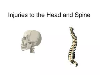
Injuries of the spine. cranial. right. posterior (dorsal). anterior (ventral). left. caudal. AP View (from posterior). Lateral View. Anatomy : Spinal Column - Orientation. Anatomy: Spinal Column - Vertebrae. Cervical Vertebrae: 7. Thoracic Vertebrae: 12. Lumbar Vertebrae: 5.

E N D
cranial right posterior (dorsal) anterior (ventral) left caudal AP View (from posterior) Lateral View Anatomy: Spinal Column - Orientation
Anatomy: Spinal Column - Vertebrae Cervical Vertebrae: 7 Thoracic Vertebrae: 12 Lumbar Vertebrae: 5 Lordosis Sacral Vertebrae: 5 Coccygeal Vertebrae: 4
Cervical Spine Anatomy C1, Atlas C2, Axis C3 C4 C5 C6 C7 A flexible group of vertebrae that support the skull Spinous Processes are Bifid Normalcurvature is lordosis
Cervical Vertebrae Atlas (C1) Axis (C2) C3-C7 Has two transverse foramen
Cervical Spine X-Ray Lateralradiograph A/Pradiograph
Thoracic Anatomy Gradual increase in size of vertebrae from top to bottom T1 T2 T3 Curvature is Kyphosis T4 T5 Articulate with ribs Rigid helps support the thorax or trunk of the body T6 T7 T8 T9 Facets are aligned horizontally T10 T11 T12
Thoracic Spine X Ray Lateralradiograph A/Pradiograph
In general a typical vertebra consists of : • large vertebral body in the front • two strong bony areas called pedicles connecting the vertebral body and the posterior arch • an arch of bony structures in the back (posterior arch) = (the spinous process). • BODY PEDICLE transverse process spinous process
Lumbar Spine Anatomy L1 L2 L3 L4 L5 Curvatureis Lordotic Vertebral body is kidney shaped in MRI Facets are aligned vertically and allow bending
Lumbar Spine X Ray Lateralradiograph A/Pradiograph
Coccyx Sacrum Sacrum lateral view median section Sacrum posterior view Triangular in shape formed by the fusion of four coccygeal vertebrae Female coccyx points inferiorly Male anteriorly S1 S2 top view S2 S3 S4 S5 Sacrum ventral view
Vertebral body Foramen Pedicle L3 medial section Processus articularis Transverse process Lamina Spinous process L4 superior view
Vertebral Body Thick disc-shaped anterior portion Weight bearing part Superior & inferior surfaces roughened for attachment of the cartilaginous intervertebral disc Anterior & lateral surfaces contain nutrient foramina for blood vessels
Anterior Posterior b a b 1 3 4 5 6 2 • Intervertebral Disc • Two Vertebrae • Ligaments • Anterior Longitudinal Ligament • Posterior Longitudinal Ligament • Capsular Ligament • Ligamentum Flavum • Interspinous Ligament • Supraspinous Ligament
Ligaments PLL ALL ALL
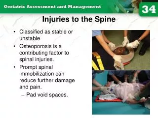
PATHOPHYSIOLOGY OF SPINE INJURIES Stable and unstable injuries Spinal injuries carry a double threat: damage to the vertebral column and damage to the neural tissues. While the full extent of the damage may be apparent from the moment of injury, there is always the fear that movement may cause or aggravate the neural lesion; hence the importance of establishing whether the injury is stable or unstable and treating it as unstable until proven otherwise. A stable injury is one in which the vertebral components will not be displaced by normal movements; in a stable injury, if the neural elements are undamaged there is little risk of them becoming damaged. An unstable injury is one in which there is a significant risk of displacement and consequent damage – or further damage – to the neural tissues.
Three column principle Posterior column Anterior column Anterior longitudinal ligament Anterior part of annulus Anterior part of vertebral body Posterior arch Posterior ligaments Inter-, Supraspinous lig. Ligamentum flavum Middle column Posterior part of vertebral body Posterior longitudinal ligament Posteror part of annulus
Pathophysiology Primary changes Physical injury may be limited to the vertebral column, including its soft-tissue components, and varies from ligamentous strains to vertebral fractures and fracture-dislocations. The spinal cord and/or nerve roots may be injured, either by the initial trauma or by ongoing structural instability of a vertebral segment, causing direct compression, severe energy transfer, physical disruption or damage to its blood supply. Secondary changes During the hours and days following a spinal injury biochemical changes may lead to more gradual cellular disruption and extension of the initial neurological damage.
Mechanism of injury Traction injury In the lumbar spine resisted muscle effort may avulse transverse processes; in the cervical spine the seventh spinous process can be avulsed (‘clay shoveller’s fracture’). Direct injury Penetrating injuries to the spine, particularly from firearms and knives, are becoming increasingly common. Indirect injury This is the most common cause of significant spinal damage; it occurs most typically in a fall from a height when the spinal column collapses in its vertical axis, NOTE: Insufficiency fractures may occur with minimal force in bone which is weakened by osteoporosis or a pathological lesion.
(a) (b) Mechanism of injury The spine is usually injured in one of two ways: (a) a fall onto the head or the back of the neck; and (b) a blow on the forehead, which forces the neck into hyperextension.
DIAGNOSIS • History. • Examination. 1.NECK; 2.BACK; The patient is ‘log-rolled’ (i.e. turned over ‘in one piece’) to avoid movement of the vertebral column. The back is inspected for deformity
3. GENERAL EXAMINATION – ‘SHOCK. 4. NEUROLOGICAL EXAMINATION. 5.IMAGING. • X-ray examination. • CT is ideal for showing structural damage to individual vertebrae. • MRI. - Remember that the spine may be damaged in more than one place. - Do not accept poor quality images. - Consult with the radiologist.
CERVICAL SPINE INJURIES The patient will usually give a history of a fall from a height, a diving accident or a vehicle accident in which the neck is forcibly moved. In a patient unconscious from a head injury, a fractured cervical spine should be assumed (and acted upon) until proved otherwise. An abnormal position of the neck is suggestive, and careful palpation may elicit tenderness.
UPPER CERVICAL SPINE Occipital condyle fracture This is usually a high-energy fracture and associated skull or cervical spine injuries must be sought. The diagnosis is likely to be missed on plain x-ray examination and CT is essential. Impacted and undisplaced fractures can be treated by brace immobilization for 8–12 weeks. Displaced fractures are best managed by using a halo-vest or by operative fixation.
C1 ring fracture Sudden severe load on the top of the head may cause a ‘bursting’ force which fractures the ring of the atlas (Jefferson’s fracture). There is no encroachment on the neural canal and, usually, no neurological damage. The fracture is seen on the open-mouth view (if the lateral masses are spread away from the odontoid peg) and the lateral view. A CT scan is particularly helpful in defining the fracture.
Fracture of C1 ring Jefferson’s fracture – bursting apart of the lateral masses of C1.
Treatment if stable and the patient wears • a semi-rigid collar • Or halo-vest until the fracture unites. If there is sideways spreading of the lateral masses (more than 7 mm on the open-mouth view), the transverse ligament has ruptured; this injury is unstable and should be treated by a halo-vest for several weeks. If there is persisting instability on x-ray, a posterior C1/2 fixation and • Fractures of the atlas are associated with injury elsewhere in the cervical spine in up to 50 per cent of cases.
C2 pars interarticularis fractures ‘hangman’s fracture’ there are • bilateral fractures of the pars interarticularis of C2 and the C2/3 disc is torn; 2. the mechanism is extension with distraction. This is one cause of death in motor vehicle accidents when the forehead strikes the dashboard. • Neurological damage, is unusual because the fracture of the posterior arch tends to decompress the spinal cord. • Nevertheless the fracture is potentially unstable.
Undisplaced fractures which are shown to be stable on supervised flexion–extension views (less than 3mm of C2/3 subluxation) can be treated in a semi-rigid orthosis until united (usually 6–12 weeks). Fractures with more than 3mm displacement but no kyphoticangulation may need reduction; however, because the mechanism of injury usually involves distraction, traction must be avoided. After reduction, the neck is held in a halo-vest until union occurs. C2/3 fusion is sometimes required for persistent pain and instability (‘traumatic spondylolisthesis’). Occasionally, the ‘hangman’s fracture’ is associated with a C2/3 facet dislocation. This is a severely unstable injury; open reduction and stabilization is required.
Fracture of C2 ‘Hangman’s fracture’ – fracture of the pars interarticularis of C2.
C2 Odontoid process fracture • uncommon. • occur as flexion injuries in young adults after highvelocity accidents or severe falls. • Also occur in elderly, osteoporotic people as a result of low-energy trauma in which the neck is forced into hyperextension, • cord damage is not uncommon and in • old people there is a considerable mortality rate
Classification D’Alonzo • Type I – An avulsion fracture of the tip of the odontoidprocessdue to traction by the alar ligaments. The fracture is stable (above the transverse ligament) and unites without difficulty. • Type II – A fracture at the junction of the odontoid process and the body of the axis. This is the most common (and potentially the most dangerous) type. The fracture is unstable and prone to non-union. • Type III – A fracture through the body of the axis. The fracture is stable and almost always unites with immobilization. (type I) (type II) (type III)
Clinical features • The history is usually that of a severe neck strain • followed by pain and stiffness due to muscle spasm. • The diagnosis is confirmed by high quality x-ray
Imaging • Plain x-rays usually show the fracture, although the • Tomography is helpful • MRIhas the advantage that it may reveal rupture of the transverse ligament; this can cause
Treatment Type I fractures Isolated fractures of the odontoid tip are uncommon. They need no more than • immobilization in a rigid collar until discomfort subsides. Type II fractures These are often unstable and prone to non-union, especially if displaced more than 5 mm. • Undisplaced fractures can be held by fitting a halo-vest • Displacedfractures should be reduced by traction and can thenbe held by operative posterior C1/2 fusion;
Type III fractures If • undisplaced, these are treated in a halo-vest for 8–12 weeks. • If displaced, attempts should be made at reducing the fracture by halo traction, which will allow positioning in either flexion or extension, depending on whether the displacement is forward or backward; the neck is then immobilized in a halo-vest for 8–12 weeks.
LOWER CERVICAL SPINE C3 to C7 • Posterior ligament injury • Sudden flexion of the mid-cervical spine in • damage to the posterior ligament complex (the interspinousligament, facet capsule and supraspinous ligament). • The upper vertebra tilts forward on the one below, opening up the interspinous space posteriorly.
The patient complains of • pain and there may be localized tenderness posteriorly • X-ray may reveal a slightly increased gap between the adjacent spines; • if the neck is held in extension this sign can be missed, • so it is always advisable to obtain a lateral view with the neck in the neutral position. A flexion view would, of course, show the widened interspinousspacethe injury is unstable and it should be treated as a subluxation or dislocation. If it is certain that the • injury is stable, a semi-rigid collar for 6 weeks is adequate; • if the injury is unstable then posterior fixation and fusion is advisable.
Wedge compression fracture • A pure flexion injury results in a wedge compression fracture of the vertebral body .The middle and posterior elements remain intact and the • injury is stable. All that is needed is a comfortable collar for 6–12 weeks. • . Diagnosis. 1. The x-ray should be carefully examined to exclude damage to the middle column and posterior displacement of the vertebral body fragment, i.e. features of a burst fracture (see below) which is potentially dangerous. If there is the least doubt 2. an axial CT 3. MRI should be obtained.
Burst and compression-flexion (‘teardrop’) fractures • These severe injuries are due to axial compression of the cervical spine, • usually in diving or athletic accidents If the vertebral body is crushed in • neutral position of the neck the result is a ‘burst • fracture’. With combined axial compression and flexion, • an antero-inferior fragment of the vertebral body is sheared off, producing the eponymous ‘tear-drop’ • on the lateral x-ray. In both types of fracture there is a risk of posterior displacement of the vertebral body fragment and spinal cord injury.
diagnosis • Plain x-rays show either a crushed vertebral body (burst fracture) or a flexion deformity with a triangular fragment separated from the antero-inferior edge of the fractured vertebra (the innocent-looking ‘teardrop’). • CT or MRI should be performed to look for retropulsion of bone fragments into the spinal canal.
TREATMENT If there is no neurological deficit, the patient can be treated surgically or by confinement to bed and traction • for 2–4 weeks, followed by a further period ofimmobilization in a halo-vest for 6–8 weeks. • If there is any deterioration of neurological status while the fracture is believed to be unstable, and the a threat of cord compression, urgent anterior decompression is considered – anterior corpectomy, bone grafting and plate fixation, and sometimes also posterior stabilization.
Fracture-dislocations • Bilateral facet joint dislocations are caused by severe flexion or flexion–rotation injuries. • The inferior articular of one facets of one vertebra ride forward over the superior facets of the vertebra below. • Diagnosis • Lat X RAY • MRI
TREATMENT • it may be more convenient to immobilize the neck in a halo-vest for 12 weeks. • Another alternative is to carry out a posterior fusion as soon as reduction has been achieved; the patient is then allowed up in a cervical brace which is worn for 6–8 weeks. • Posterior open reduction and fusion is also indicated if closed reduction fails. • Unilateral facet dislocation is the same as for bilateral dislocation. Sometimes complete reduction is prevented by the upper facet becoming perched upon the lower.