Download

1 / 64
690 likes | 1.07k Views
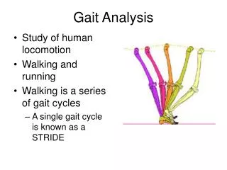
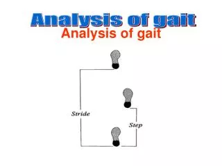
Gait Analysis: Technology and Clinical Applications. دکترامیر هوشنگ واحدی متخصص طب فیزیکی و توانبخشی قسمت 2. Fine Movements. Pelvis undergoes slight

E N D
Gait Analysis: Technology and Clinical Applications دکترامیر هوشنگ واحدی متخصص طب فیزیکی و توانبخشی قسمت 2
Fine Movements • Pelvis undergoes slight • Legsrotateslightly medially at hip andknee during swing and heel-strike tonear mid-stance, followed by a changeto lateral rotation which continuesthrough push-off. • Knees have two alternations of extension and flexion in a single cycle • Ankle
Determinants of Gait : • Six optimizations used to minimize excursion of COG in vertical & horizontal planes • Reduce significantly energy consumption of ambulation
The six determinants(optimizations )of gait are 1. Pelvic rotation 2. Pelvic tilt 3. Knee flexion in stance phase 4. Foot mechanisms 5. Knee mechanisms 6. Lateral displacement of the pelvis
(1) Pelvic rotation: Forward rotation of the pelvis in the horizontal plane approx. 8oon the swing-phase side Reduces the angle of hip flexion & extension
(2) Pelvic tilt: Reduces the height of the apex of the curve of COG The pelvis on the side of the swinging leg (opposite to the weight-bearing leg) is lowered 4°–5°, which lowers COG at midstance
(3) Knee flexion in stance phase: Approx. 15o Shortens the leg in the middle of stance phase Reduces the height of the apex of the curve of COG
(4) Ankle mechanism: Lengthens the leg at heel contact Smoothens the curve of COG Reduces the lowering of COG
5) Foot mechanism: Lengthens the leg at toe-off as ankle moves from dorsiflexion to plantarflexion Smoothens the curve of COG Reduces the lowering of COG Late knee flexion(30-40")duringthe last part of the stance phase.
(6) Lateral displacement of body: The normally narrow width of the walking base minimizes the lateral displacement of COG Reduced muscular energy consumption due to reduced lateral acceleration & deceleration
Determinants 1–5 reduce displacement on the vertical plane. • Determinant 6 reduces displacement on the horizontal plane.
What is Gait Analysis? An objective measurement of human locomotion
Instrumented gait analysis Analysis includes the following. • Video with slow motion and superimposed force line visualization. • Poly-EMG • Kinematic temporal-spatial parameters of locomotion. • Kinetic analysis to quantify joint moments and powers.
Force Platforms Baylor Motion & Sports Performance Center • Vicon Motion Analysis System • Twelve MX-40 Cameras • 4 – Megapixel Resolution • 5 Hz – 2,000 Hz • Passive Marker System • Near Infra-Red Ring LEDs • Camera/Computer
Direct Linear Transformation Virtual Hip Joint Centers Camera 1 Camera 2 Identified Markers 3D Image 3D Image DLT Camera 3………… Camera 12 Take all 12 Raw Video Camera Views
Four primary components of quantitative gait analysis that can be recorded are: I. kinetics (analysis of forces that produce motion); 2. poly-EMG or dynamic EMG (analysis of muscle activity); 3. kinematics (analysis of motion and resulting temporal and stride measures) 4. energetics(analysis of metabolic or mechanical energy).
Newton described basic but critical concepts that are useful in understanding the effect of gravity on gait. • The ground reaction force is a reflection of the body weight and the acceleration of our body. • Kinetics involves measurement of joint forces and moments
Kinematics - the movement of the body in space without any reference to forces.
Dynamic polyelectromyography • In normal locomotion, forces are elicited from 28 muscles in each lower limb to carefully control the gravitational forces, coordinated, and energy-efficient movement pattern. • Measures electrical activity of a contracting muscle • Assesses timing, relative intensity and absence of activity
Energetics • Normal walking requires a relatively low level of metabolic energy consumption during steady state at comfortable walking speeds. • Normal gait on level surfaces is most efficient at a waking speed of 1-1.3 m/s, which is equivalent to 60-80 m/ min or 3 miles/h. • Comfortable walking speed for an individual usually corresponds to minimum energy cost per unit distance.
There are several methods of metabolic energy measurement. • Indirect calorimetry • expired air collection • heart rate monitoring
Assessing Oxygen Consumption Physiological cost index (PCI) Heart rate and walking speed O2 consumption and/or CO2 production
There is a link between motion of the COM and energy expended during walking. • Sudden acceleration or deceleration of the COM will increase energy consumption. • Running is more efficient than walking faster than 2 m/s • Walking on a 10-12% incline will double energy expenditure.
The three main events that consume energy during walking are • controlled deceleration toward the end of swing phase, • shock absorption at heel strike • forward propulsion of the COM at push-off.
Posterior and lateral views of the major muscles of the lower extremities, showing the activity of the muscles at key phases of the gait cycle. 1.gluteus maximus (posterior view); 2. gluteus medius (posterior and lateral views); 3. adductor magnus (posterior view); 4. quadriceps (lateral view); 5. hamstrings (posterior and lateral views); 6. tibialis anterior (lateral view). 7.iliopsoas
Initial Contact : • Knee Biomechanics: The knee is positioned in neutral The quadriceps contract to prepare for loading response • Hip Biomechanics: Hip stays in the 25 degrees of flexion obtained in terminal swing. Hamstrings contract in reaction to hip flexion torque.
Loading Response : • Knee Biomechanics: The knee flexes to 15 degrees Quadriceps contract eccentrically. • Hip Biomechanics: Hip remains in 25 degrees of flexion. Glut max, hamstrings, and adductors contract
Midstance : • Knee Biomechanics: The knee extends near neutral. then the knee is stabilized by the gastrocnemius. • Hip Biomechanics: Hip extends to neutral. No muscle activity in the sagittal plane. The pelvis is stabilized in the frontal plane by the hip abductor group.
Terminal Stance : • Knee Biomechanics: Gait Terminal Stance The knee extends to neutral. No muslces are active at the knee. Knee still stabilized by the gastrocs. • Hip Biomechanics: Gait Terminal Stance Hip extends 20 degrees.
PreSwing : • Knee Biomechanics: The knee rapidly flexes to 40 degrees. The motion occurs without hip flexor activity. • Hip Biomechanics: Thigh falls forward. This motion is aided by adductor longus.
Initial Swing • Knee Biomechanics: The knee continues flexing to 60 degrees. Biceps femoris, sartorius, and gracilis are active. Foot clears the floor and the thigh advances. • Hip Biomechanics: Gait Initial Swing 15 degrees of hip flexion is achieved, Iliacus, gracilis, sartorius, and adductor longus are active concentrically.