Download
1 / 57
610 likes | 766 Views
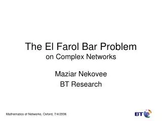
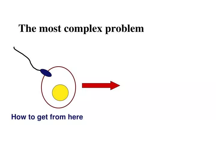
The most complex problem. How to get from here. The most complex problem. How to get from here to there . Chapter 47. Animal Development. Figure 47.1. A human embryo at about 7 weeks after conception shows development of distinctive features. 1 mm. Development: cellular level .

E N D
The most complex problem How to get from here
The most complex problem How to get from here to there
Chapter 47 Animal Development
Figure 47.1 A human embryo at about 7 weeks after conception shows development of distinctive features 1 mm
Development: cellular level • Cell division • Differentiation • cells become specialized in structure & function • Morphogenesis (organogenesis)
Development: cellular level • Cell division • Differentiation • cells become specialized in structure & function • if each kind of cell has the same genes, how can they be so different? • shutting off of genes = loss of totipotency • Turning genes on based on chemical cues • Morphogenesis (organogenesis)
Development: cellular level • Cell division • Differentiation • cells become specialized in structure & function • if each kind of cell has the same genes, how can they be so different? • shutting off of genes = loss of totipotency • Turning genes on based on chemical cues • Morphogenesis (organogenesis) • “creation of form” = give organism shape • basic body plan • polarity • one end is different than the other • symmetry • left & right side of body mirror each other • asymmetry • look at your hand…
EMBRYONIC DEVELOPMENT Developmental events Sperm Zygote Adultfrog Egg FERTILIZATION CLEAVAGE Metamorphosis Blastula GASTRULATION ORGANO-GENESIS Larvalstages Gastrula Tail-budembryo
Fertilization in sea urchins: fast block and slow block to polyspermy Basal body(centriole) Spermhead Acrosome Jelly coat Vitelline layer Sperm-bindingreceptors Egg plasma membrane
Figure 47.3-2 Basal body(centriole) Spermhead Acrosome Hydrolytic enzymes Jelly coat Vitelline layer Sperm-bindingreceptors Egg plasma membrane
Figure 47.3-3 Spermnucleus Acrosomalprocess Basal body(centriole) Actinfilament Spermhead Acrosome Hydrolytic enzymes Jelly coat Vitelline layer Sperm-bindingreceptors Egg plasma membrane
Figure 47.3-4 Spermplasmamembrane Spermnucleus Acrosomalprocess Basal body(centriole) Actinfilament Spermhead Fusedplasmamembranes Acrosome Hydrolytic enzymes Jelly coat Vitelline layer Sperm-bindingreceptors Egg plasma membrane
Figure 47.3-5 Spermplasmamembrane Spermnucleus Fertilizationenvelope Acrosomalprocess Basal body(centriole) Actinfilament Spermhead Corticalgranule Fusedplasmamembranes Acrosome Hydrolytic enzymes Perivitellinespace Jelly coat Vitelline layer Sperm-bindingreceptors EGG CYTOPLASM Egg plasma membrane
EXPERIMENT Slow block to polyspermy: Change in Ca++ in the egg makes f.e. 25 sec 10 sec afterfertilization 1 min 35 sec 500 m RESULTS 1 sec beforefertilization 10 sec afterfertilization 30 sec 20 sec 500 m CONCLUSION Fertilizationenvelope Spreadingwave of Ca2 Point of spermnucleusentry
© 2011 Pearson Education, Inc. Egg Activation • The rise in Ca2+ in the cytosol increases the rates of cellular respiration and protein synthesis by the egg cell • With these rapid changes in metabolism, the egg is said to be activated • The proteins and mRNAs needed for activation are already present in the egg • The sperm nucleus merges with the egg nucleus and cell division begins
When the sperm binds a receptor in the zona pellucida, it triggers a slow block to polyspermy (no fast block to polyspermy has been identified in mammals) Zona pellucida Follicle cell Corticalgranules Spermnucleus Spermbasal body
CLEAVAGE • Fertilization is followed by cleavage, a period of rapid cell division without growth • Cleavage partitions the cytoplasm of one large cell into many smaller cells called blastomeres • The blastula is a ball of cells with a fluid-filled cavity called a blastocoel 50 m (d) Later blastula (a) Fertilized egg (c) Early blastula (b) Four-cell stage
Zygote Cleavage in a frog embryo 2-cellstageforming Gray crescent 0.25 mm 8-cell stage (viewedfrom the animal pole) 4-cellstageforming Animalpole 8-cellstage 0.25 mm Blastula (at least 128 cells) Vegetal pole Blastocoel Blastula(crosssection)
© 2011 Pearson Education, Inc. Concept 47.2: Morphogenesis in animals involves specific changes in cell shape, position, and survival • Morphogenesis, the process by which cells occupy their appropriate locations, involves • Gastrulation, the movement of cells from the blastula surface to the interior of the embryo • Organogenesis, the formation of organs
ECTODERM (outer layer of embryo) Figure 47.8 • Epidermis of skin and its derivatives (including sweat glands, hair follicles) • Nervous and sensory systems • Pituitary gland, adrenal medulla • Jaws and teeth • Germ cells MESODERM (middle layer of embryo) • Skeletal and muscular systems • Circulatory and lymphatic systems • Excretory and reproductive systems (except germ cells) • Dermis of skin • Adrenal cortex ENDODERM (inner layer of embryo) • Epithelial lining of digestive tract and associated organs (liver, pancreas) • Epithelial lining of respiratory, excretory, and reproductive tracts and ducts • Thymus, thyroid, and parathyroid glands
Animalpole Blastocoel Gastrulation in the sea urchin Mesenchymecells Vegetal plate Vegetalpole Blastocoel Filopodia Mesenchymecells Archenteron Blastopore 50 m Blastocoel Ectoderm Archenteron Blastopore Key Mouth Future ectoderm Mesenchyme(mesoderm formsfuture skeleton) Digestive tube (endoderm) Future mesoderm Anus (from blastopore) Future endoderm
2 3 1 CROSS SECTION SURFACE VIEW Animal pole Gastrulation in frog embryo Blastocoel Dorsal lip ofblasto-pore Dorsal lip ofblastopore Blastopore Earlygastrula Vegetal pole Blastocoelshrinking Archenteron Ectoderm Blastocoelremnant Mesoderm Endoderm Key Future ectoderm Blastopore Lategastrula Future mesoderm Yolk plug Archenteron Blastopore Future endoderm
Fertilized egg Primitivestreak Gastrulation in chicks Embryo Yolk Primitive streak Epiblast Future ectoderm Blastocoel Endoderm Migratingcells(mesoderm) Hypoblast YOLK
1 3 4 2 Endometrial epithelium(uterine lining) Blastocyst reaches uterus. Inner cell mass Uterus Embryonic development in humans Trophoblast Blastocoel Blastocyst implants(7 days after fertilization). Expanding region oftrophoblast Maternal bloodvessel Epiblast Hypoblast Trophoblast Expanding region oftrophoblast Extraembryonic membranesstart to form (10–11 days),and gastrulation begins(13 days). Amniotic cavity Epiblast Hypoblast Yolk sac (from hypoblast) Extraembryonic mesoderm cells(from epiblast) Chorion (from trophoblast) Gastrulation has produced athree-layered embryo withfour extraembryonicmembranes. Amnion Chorion Ectoderm Mesoderm Endoderm Yolk sac Extraembryonic mesoderm Allantois
1 Figure 47.12a Endometrial epithelium(uterine lining) Inner cell mass Uterus Trophoblast Blastocoel Blastocyst reaches uterus.
2 Figure 47.12b Expanding region oftrophoblast Maternal bloodvessel Epiblast Hypoblast Trophoblast Blastocyst implants(7 days after fertilization).
3 Figure 47.12c Expanding region oftrophoblast Amniotic cavity Epiblast Hypoblast Yolk sac (from hypoblast) Extraembryonic mesoderm cells (from epiblast) Chorion (from trophoblast) Extraembryonic membranesstart to form (10–11 days),and gastrulation begins(13 days).
4 Figure 47.12d Amnion Chorion Ectoderm Mesoderm Endoderm Yolk sac Extraembryonic mesoderm Allantois Gastrulation has produced athree-layered embryo withfour extraembryonicmembranes.
© 2011 Pearson Education, Inc. Developmental Adaptations of Amniotes • The colonization of land by vertebrates was made possible only after the evolution of • The shelled egg of birds and other reptiles as well as monotremes (egg-laying mammals) • The uterus of marsupial and eutherian mammals
© 2011 Pearson Education, Inc. • The four extraembryonic membranesthatform around the embryo in a reptile/bird: • The chorionfunctions in gas exchange • The amnionencloses the amniotic fluid • The yolk sacencloses the yolk • The allantoisdisposes of waste products and contributes to gas exchange
Eye Somites Tail bud Neural folds Neuralation in a frog embryo Neural fold Neural plate SEM 1 mm 1 mm Neural tube Neuralcrestcells Neural plate Neural fold Notochord Coelom Neural crest cells Notochord Somite Ectoderm Outer layerof ectoderm Mesoderm Archenteron(digestivecavity) Neural crest cells Endoderm (c) Somites Archenteron (a) Neural plate formation Neural tube (b) Neural tube formation
Organogenisis in a chick Neural tube Eye Notochord Forebrain Somite Archenteron Coelom Heart Lateral fold Endoderm Bloodvessels Mesoderm Ectoderm Somites Yolk stalk Yolk sac These layersform extraembryonicmembranes. Neuraltube YOLK (a) Early organogenesis (b) Late organogenesis
Ectoderm Morpho-genesis results from cells changing shape
Ectoderm Figure 47.15-2 Neuralplate Microtubules
Ectoderm Figure 47.15-3 Neuralplate Microtubules Actinfilaments
Ectoderm Figure 47.15-4 Neuralplate Microtubules Actinfilaments
Ectoderm Figure 47.15-5 Neuralplate Microtubules Actinfilaments Neural tube
Elongation of tissue by convergent extension Convergence Extension
© 2011 Pearson Education, Inc. Concept 47.3: What determines how parts form; and messing with those determining factors • Determination is the term used to describe the process by which a cell or group of cells becomes committed to a particular fate • Differentiation refers to the resulting specialization in structure and function • Can result from: oocyte composition, logal signals, gravity
Epidermis Epidermis Centralnervoussystem Fate mapping Notochord Mesoderm Endoderm Blastula Neural tube stage(transverse section) (a) Fate map of a frog embryo 64-cell embryos Blastomeresinjected with dye Larvae (b) Cell lineage analysis in a tunicate
Zygote 0 First cell division Figure 47.18 Muscula-ture, gonads Outer skin,nervous system Germ line(futuregametes) Nervoussystem,outer skin,muscula-ture Time after fertilization (hours) Musculature 10 Hatching Intestine Intestine Anus Mouth Eggs Vulva POSTERIOR ANTERIOR 1.2 mm
2 1a 1b EXPERIMENT Control egg(dorsal view) Experimental egg(side view) How does distribution of the gray crescent affect the developmental potential of the first two daughter cells? Experimentalgroup Controlgroup Graycrescent Graycrescent Thread RESULTS Normal Belly piece Normal
Figure 47.7b 0.25 mm Animalpole 8-cell stage (viewedfrom the animal pole)
Figure 47.7c 0.25 mm Blastocoel Blastula (at least 128 cells)
Can the dorsal lip of the blastopore induce cells in another part of the amphibian embryo to change their developmental fate? RESULTS EXPERIMENT Primary embryo Dorsal lip ofblastopore Secondary (induced) embryo Pigmentedgastrula(donor embryo) Primary structures: Neural tube Nonpigmentedgastrula(recipient embryo) Notochord Secondary structures: Notochord (pigmented cells) Neural tube(mostly nonpigmented cells)
Figure 47.24 Anterior Limb bud AER ZPA Limb buds 2 Posterior 50 m Digits Apicalectodermalridge (AER) 3 4 Anterior Ventral Proximal Distal Dorsal Posterior (a) Organizer regions (b) Wing of chick embryo
© 2011 Pearson Education, Inc. • One limb bud–regulating region is the apical ectodermal ridge (AER) • The AER is thickened ectoderm at the bud’s tip • The second region is the zone of polarizing activity (ZPA) • The ZPA is mesodermal tissue under the ectoderm where the posterior side of the bud is attached to the body
EXPERIMENT Anterior New ZPA Donorlimbbud Hostlimbbud ZPA Posterior RESULTS What happens when you put ZPA on both sides of a budding limb?