Download
1 / 45
450 likes | 544 Views
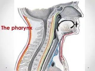
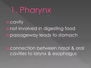
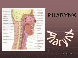
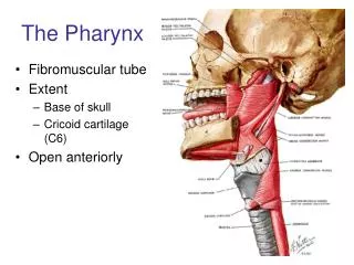
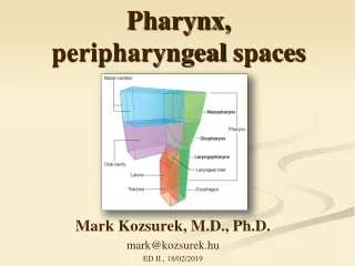
Pharynx. From the mouth, the oro- and laryngopharynx allow passage of: Food and fluids to the esophagus Air to the trachea Lined with stratified squamous epithelium and mucus glands Has two skeletal muscle layers Inner longitudinal Outer pharyngeal constrictors. Esophagus.

E N D
Pharynx • From the mouth, the oro- and laryngopharynx allow passage of: • Food and fluids to the esophagus • Air to the trachea • Lined with stratified squamous epithelium and mucus glands • Has two skeletal muscle layers • Inner longitudinal • Outer pharyngeal constrictors
Esophagus • Muscular tube going from the laryngopharynx to the stomach • Travels through the mediastinum and pierces the diaphragm • Joins the stomach at the cardiac orifice
Esophageal Characteristics • Esophageal mucosa – nonkeratinized stratified squamous epithelium • The empty esophagus is folded longitudinally and flattens when food is present • Glands secrete mucus as a bolus (compacted food product) moves through the esophagus • Muscularis changes from skeletal (superiorly) to smooth muscle (inferiorly)
Digestive Processes in the Mouth • Food is ingested • Mechanical digestion begins (chewing) • Propulsion is initiated by swallowing • Salivary amylase begins chemical breakdown of starch • The pharynx and esophagus serve as conduits to pass food from the mouth to the stomach
Deglutition (Swallowing) • Coordinated activity of the tongue, soft palate, pharynx, esophagus, and 22 separate muscle groups • Buccal phase – bolus is forced into the oropharynx
Deglutition (Swallowing) Pharyngeal-esophageal phase – controlled by the medulla and lower pons • All routes except into the digestive tract are sealed off • Peristalsis moves food through the pharynx to the esophagus
Stomach • Chemical breakdown of proteins begins and food is converted to chyme • Cardiac region – surrounds the cardiac orifice • Fundus – dome-shaped region beneath the diaphragm • Body – midportion of the stomach • Pyloric region – made up of the antrum and canal which terminates at the pylorus • The pylorus is continuous with the duodenum through the pyloric sphincter
Stomach • Greater curvature – entire extent of the convex lateral surface • Lesser curvature – concave medial surface • Lesser omentum – runs from the liver to the lesser curvature • Greater omentum – drapes inferiorly from the greater curvature to the small intestine
Stomach • Nerve supply – sympathetic and parasympathetic fibers of the autonomic nervous system • Blood supply – celiac trunk, and corresponding veins (part of the hepatic portal system)
Microscopic Anatomy of the Stomach • Muscularis – has an additional oblique layer that: • Allows the stomach to churn, mix, and pummel food physically • Breaks down food into smaller fragments
Microscopic Anatomy of the Stomach • Epithelial lining is composed of: • Goblet cells that produce a coat of alkaline mucus • The mucous surface layer traps a bicarbonate-rich fluid beneath it • Gastric pits contain gastric glands that secrete gastric juice, mucus, and gastrin
Glands of the Stomach Fundus and Body • Gastric glands of the fundus and body have a variety of secretory cells • Mucous neck cells – secrete acid mucus • Parietal cells – secrete HCl and intrinsic factor
Glands of the Stomach Fundus and Body • Chief cells – produce pepsinogen • Pepsinogen is activated to pepsin by: • HCl in the stomach • Pepsin itself via a positive feedback mechanism • Enteroendocrine cells – secrete gastrin, histamine, endorphins, serotonin, cholecystokinin (CCK), and somatostatin into the lamina propria
Stomach Lining • The stomach is exposed to the harshest conditions in the digestive tract • To keep from digesting itself, the stomach has a mucosal barrier with: • A thick coat of bicarbonate-rich mucus on the stomach wall • Epithelial cells that are joined by tight junctions • Gastric glands that have cells impermeable to HCl • Damaged epithelial cells are quickly replaced
Digestion in the Stomach • The stomach: • Holds ingested food • Degrades this food both physically and chemically • Delivers chyme to the small intestine • Enzymatically digests proteins with pepsin • Secretes intrinsic factor required for absorption of vitamin B12
Regulation of Gastric Secretion • Neural and hormonal mechanisms regulate the release of gastric juice • Stimulatory and inhibitory events occur in three phases • Cephalic (reflex) phase: prior to food entry • Gastric phase: once food enters the stomach • Intestinal phase: as partially digested food enters the duodenum
Cephalic Phase • Excitatory events include: • Sight or thought of food • Stimulation of taste or smell receptors • Inhibitory events include: • Loss of appetite or depression • Decrease in stimulation of the parasympathetic division
Gastric Phase • Excitatory events include: • Stomach distension • Activation of stretch receptors (neural activation) • Activation of chemoreceptors by peptides, caffeine, and rising pH • Release of gastrin to the blood
Gastric Phase • Inhibitory events include: • A pH lower than 2 • Emotional upset that overrides the parasympathetic division
Intestinal Phase • Excitatory phase – low pH; partially digested food enters the duodenum and encourages gastric gland activity • Inhibitory phase – distension of duodenum, presence of fatty, acidic, or hypertonic chyme, and/or irritants in the duodenum • Initiates inhibition of local reflexes and vagal nuclei • Closes the pyloric sphincter • Releases enterogastrones that inhibit gastric secretion
Regulation and Mechanism of HCl Secretion • HCl secretion is stimulated by ACh, histamine, and gastrin through second-messenger systems • Antihistamines block H2 receptors and decrease HCl release
Response of the Stomach to Filling • Stomach pressure remains constant until about 1L of food is ingested • Relative unchanging pressure results from reflex-mediated relaxation and plasticity
Response of the Stomach to Filling • Reflex-mediated events include: • Receptive relaxation – as food travels in the esophagus, stomach muscles relax • Adaptive relaxation – the stomach dilates in response to gastric filling • Plasticity – intrinsic ability of smooth muscle to exhibit the stress-relaxation response
Gastric Contractile Activity • Peristaltic waves move toward the pylorus at the rate of 3 per minute • This basic electrical rhythm (BER) is initiated by pacemaker cells (cells of Cajal)
Gastric Contractile Activity • Most vigorous peristalsis and mixing occurs near the pylorus • Chyme is either: • Delivered in small amounts to the duodenum or • Forced backward into the stomach for further mixing
Regulation of Gastric Emptying • Gastric emptying is regulated by: • The neural enterogastric reflex • Hormonal (enterogastrone) mechanisms • These mechanisms inhibit gastric secretion and duodenal filling
Regulation of Gastric Emptying • Carbohydrate-rich chyme quickly moves through the duodenum • Fat-laden chyme is digested more slowly causing food to remain in the stomach longer
Small Intestine: Gross Anatomy • Runs from pyloric sphincter to the ileocecal valve • Has three subdivisions: duodenum, jejunum, and ileum
Small Intestine: Gross Anatomy • The bile duct and main pancreatic duct: • Join the duodenum at the hepatopancreatic ampulla • Are controlled by the sphincter of Oddi • The jejunum extends from the duodenum to the ileum • The ileum joins the large intestine at the ileocecal valve
Small Intestine: Microscopic Anatomy • Structural modifications of the small intestine wall increase surface area • Plicae circulares: deep circular folds of the mucosa and submucosa • Villi – fingerlike extensions of the mucosa • Microvilli – tiny projections of absorptive mucosal cells’ plasma membranes
Small Intestine: Histology of the Wall • The epithelium of the mucosa is made up of: • Absorptive cells and goblet cells • Enteroendocrine cells • Interspersed T cells called intraepithelial lymphocytes (IELs) • IELs release cytokines
Small Intestine: Histology of the Wall • Cells of intestinal crypts secrete intestinal juice • Peyer’s patches are found in the submucosa • Brunner’s glands in the duodenum secrete alkaline mucus
Intestinal Juice • Secreted by intestinal glands in response to distension or irritation of the mucosa • Slightly alkaline and isotonic with blood plasma • Largely water, enzyme-poor, but contains mucus
Liver • The largest gland in the body • Superficially has four lobes – right, left, caudate, and quadrate • The falciform ligament: • Separates the right and left lobes anteriorly • Suspends the liver from the diaphragm and anterior abdominal wall
Liver • The ligamentum teres: • Is a remnant of the fetal umbilical vein • Runs along the free edge of the falciform ligament
Liver: Associated Structures • The lesser omentum anchors the liver to the stomach • The hepatic blood vessels enter the liver at the porta hepatis • The gallbladder rests in a recess on the inferior surface of the right lobe
Liver: Associated Structures • Bile leaves the liver via: • Bile ducts, which fuse into the common hepatic duct • The common hepatic duct, which fuses with the cystic duct • These two ducts form the bile duct
Liver: Microscopic Anatomy • Hexagonal-shaped liver lobules are the structural and functional units of the liver • Composed of hepatocyte (liver cell) plates radiating outward from a central vein • Portal triads are found at each of the six corners of each liver lobule Figure 23.24c
Liver: Microscopic Anatomy • Portal triads consist of a bile duct and • Hepatic artery – supplies oxygen-rich blood to the liver • Hepatic portal vein – carries venous blood with nutrients from digestive viscera Figure 23.24d
Liver: Microscopic Anatomy • Liver sinusoids – enlarged, leaky capillaries located between hepatic plates • Kupffer cells – hepatic macrophages found in liver sinusoids
Liver: Microscopic Anatomy • Hepatocytes’ functions include: • Production of bile • Processing bloodborne nutrients • Storage of fat-soluble vitamins • Detoxification • Secreted bile flows between hepatocytes toward the bile ducts in the portal triads
Composition of Bile • A yellow-green, alkaline solution containing bile salts, bile pigments, cholesterol, neutral fats, phospholipids, and electrolytes • Bile salts are cholesterol derivatives that: • Emulsify fat • Facilitate fat and cholesterol absorption • Help solubilize cholesterol • Enterohepatic circulation recycles bile salts • The chief bile pigment is bilirubin, a waste product of heme
The Gallbladder • Thin-walled, green muscular sac on the ventral surface of the liver • Stores and concentrates bile by absorbing its water and ions • Releases bile via the cystic duct, which flows into the bile duct
Regulation of Bile Release • Acidic, fatty chyme causes the duodenum to release: • Cholecystokinin (CCK) and secretin into the bloodstream • Bile salts and secretin transported in blood stimulate the liver to produce bile • Vagal stimulation causes weak contractions of the gallbladder
Regulation of Bile Release • Cholecystokinin causes: • The gallbladder to contract • The hepatopancreatic sphincter to relax • As a result, bile enters the duodenum