Download
1 / 89
900 likes | 913 Views
Microscopy and Cell Structure. Chapter 3 Part I Observing Cells. Microscope Techniques Microscopes. Principles of Light Microscopy. Light Microscopy Most common and easiest to use: bright-field microscope Important factors in light microscopy include Magnification Resolution Contrast.

E N D
Microscopy and Cell Structure Chapter 3 Part I Observing Cells
Principles of Light Microscopy • Light Microscopy • Most common and easiest to use: bright-field microscope • Important factors in light microscopy include • Magnification • Resolution • Contrast
Principles of Light Microscopy • Magnification • two magnifying lenses • Ocular lens and objective lens • condenser lens • focus illumination on specimen
Principles of Light Microscopy • Resolution • minimum distance between two objects that still appear as separate objects • determine the usefulness of microscope
Principles of Light Microscopy • Factors affect resolution • Lens • Wavelength of light • How much light is released from the lens • magnification • Maximum resolving power of most brightfield microscopes is 0.2 μm (1x10⁻⁶) • sufficient to see most bacteria • Too low to see viruses
Principles of Light Microscopy • Resolution is enhanced with lenses of higher magnification (100x) by the use of immersion oil • Oil reduces light refraction • Immersion oil has nearly same refractive index as glass
Principles of Light Microscopy • Contrast • Reflects the number of visible shades in a specimen • increase contrast • Use special microscopes • specimen staining
Principles of Light Microscopy • Examples of light microscopes that increase contrast • Phase-Contrast Microscope • Interference Microscope • Dark-Field Microscope • Fluorescence Microscope • Confocal Scanning Laser Microscope
Principles of Light Microscopy • Phase-Contrast • Amplifies differences between refractive indexes of cells and surrounding medium • Darker appearance for denser materials. • Uses set of rings and diaphragms to achieve resolution
Principles of Light Microscopy • Interference Scope • appear three dimensional • Depends on differences in refractive index
Principles of Light Microscopy • Dark-Field Microscope • Reverse image • Like a photographic negative • a modified condenser directs the lights at an angle and only the light scattered by the specimen enters the objective lens
Principles of Light Microscopy • Fluorescence Microscope • observe organisms naturally fluorescent or flagged with fluorescent dye • Fluorescent molecule absorbs ultraviolet light and emits visible light • Image fluoresces on dark background
Principles of Light Microscopy • Electron Microscope • Uses electromagnetic lenses, electrons and fluorescent screen to produce image • Resolution increased 1,000 fold over brightfield microscope • To about 0.3 nm (1x10⁻⁹) • Magnification increased to 100,000x • Two types of electron microscopes • Transmission • Scanning
Quiz • With 10x ocular lens and 40x objective lens, what is the magnifying power?
Quiz • What are the three important factors for microscope?
Microscope TechniquesDyes and Staining • Dyes and Staining • stained to observe organisms • made of organic salts • Basic dyes carry positive charge • Acidic dyes carry negative charge
Microscope TechniquesDyes and Staining • Common basic dyes include • Methylene blue • Crystal violet • Safrinin • Malachite green
Microscope TechniquesDyes and Staining • Simple staining • use one color to stain • increase contrast between cell and background
Microscope TechniquesDyes and Staining • Differential Stains • to distinguish one bacterial group from another • Uses a series of reagents • Two most common differential stains • Gram stain • Acid-fast stain
Microscope TechniquesDyes and Staining • Gram Stain • widely used procedure for classiffying bacteria • two major groups based on cell wall structural differences • Gram positive • Gram negative
Microscope TechniquesDyes and Staining • Gram Stain • Involves four reagents • Primary stain • Mordent • Decolorizer • Counter or Secondary stain Old gram positive appears to be gram negative
Microscope TechniquesDyes and Staining • Acid-fast Stain • Used to stain members of genus Mycobacterium • High lipid concentration in cell wall • Uses heat to facilitate staining
Microscope TechniquesDyes and Staining • Acid-fast Stain • used for presumptive identification in diagnosis of clinical specimens • Requires multiple steps • Primary dye • Decolorizer • Counter stain
Microscope TechniquesDyes and Staining • Special Stains • Capsule stain • Endospore stain • Uses heat to facilitate staining • Flagella stain
Quiz • What are the two commonly used differential staining method?
Morphology of Prokaryotic Cells • Prokaryotes exhibit a variety of shapes • Coccus • Bacillus • Do not to be confused with Bacillus genus
Morphology of Prokaryotic Cells • Coccobacillus • Vibrio • Spirillum • Spirochete • Pleomorphic
Morphology of Prokaryotic Cells • groupings morphology • Cells adhere together after cell division for characteristic arrangements • Especially in the cocci
Morphology of Prokaryotic Cells • Division along a single plane may result in pairs or chains of cells • Pairs = diplococci • Example: Neisseria gonorrhoeae • Chains = streptococci • Example: species of Streptococcus
Morphology of Prokaryotic Cells • Division along two or three perpendicular planes form cubical packets • Example: Sarcina genus • Division along several random planes form clusters • Example: species of Staphylococcus
Microscope • Three important factors • Staining. • Prokaryotic morphology
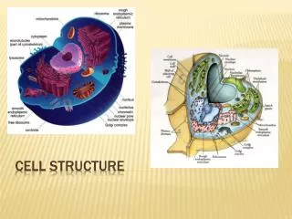
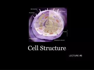
Microscopy and Cell Structure Part II - Prokaryotic Cell Structure
Cytoplasmic membrane • Defines the boundary of the cell • Semi-permeable; • Transport proteins function as selective gates (selectively permeable) • Control entrance/expulsion of antimicrobial drugs • Receptors provide a sensor system • Phospholipid bilayer, embedded with proteins
Cytoplasmic membrane • Defines the boundary of the cell • Semi-permeable; • Transport proteins function as selective gates (selectively permeable) • Control entrance/expulsion of antimicrobial drugs • Receptors provide a sensor system • Phospholipid bilayer, embedded with proteins
Cytoplasmic membrane • Defines the boundary of the cell • Semi-permeable; • Transport proteins function as selective gates (selectively permeable) • Control entrance/expulsion of antimicrobial drugs • Receptors provide a sensor system • Phospholipid bilayer, embedded with proteins • Fluid mosaic model
Cytoplasmic Membrane • Methods for molecule to go cross membrane • Simple diffusion: the only system does not rely on transport protein • Facilitated diffusion • Active transport • Group transport
Cytoplasmic Membrane • Simple diffusion- • Water, certain gases and small hydrophobic molecules • Move along with concentration gradient • Osmosis
Cytoplasmic Membrane • Movement of molecules across membrane by transport systems • Specific Transport systems include • Facilitated diffusion • Active transport • Group translocation
Directed Movement of Molecules Across the Cytoplasmic Membrane Facilitated diffusion no energy expended
Directed Movement of Molecules Across the Cytoplasmic Membrane Facilitated diffusion Active transport - energy is expended • Moves compounds against a concentration gradient
Major facilitator superfamily (expends proton motive force) ABC transport systems (expends ATP) Directed Movement of Molecules Across the Cytoplasmic Membrane Facilitated diffusion Active transport - energy is expended • Use binding proteins to scavenge and deliver molecules to transport complex • Example: maltose transport • Example: efflux pumps used in antimicrobial resistance
Cytoplasmic membrane Proton: H+ Proton motive force: Energy stored in the electrochemical gradient created by electron transport chain Electron transport chain Electron transport chain - Series of proteins that sequentially transfer electrons and eject protons from the cell, creating an electrochemical gradient • Proton motive force is used to fuel: • Synthesis of ATP (the cell’s energy currency) • Rotation of flagella (motility) • One form of transport
Directed Movement of Molecules Across the Cytoplasmic Membrane Facilitated diffusion Active transport - Chemically modifies a compound during transport Group translocation
Directed Movement of Molecules Across the Cytoplasmic Membrane Facilitated diffusion Active transport Group translocation Secretion - Transport of proteins to the outside • Characteristic sequence of amino acids in a newly synthesized protein functions as a tag (signal sequence)
Prokaryotic structure • Cell membrane structure • Movements across membrane
Cell Wall Provides rigidity to the cell (prevents it from bursting)
Cell Wall • Bacterial cell wall • Rigid structure • Determines shape of bacteria • Protection • Unique chemical structure • Distinguishes Gram positive from Gram-negative
Cell Wall • Peptidoglycan- rigid molecule; unique to bacteria • Alternating subunits of NAG and NAM form glycan chains • Glycan chains are connected to each other via peptide chains on NAM molecules