Download
1 / 8
180 likes | 563 Views
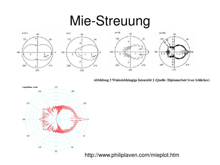
Mie-Streuung. http://www.philiplaven.com/mieplot.htm. Mie – Rayleigh-Debye-Gans. Mie: 2a=1000nm 2a=300nm 2a=200nm 2a=20nm. n solvent =1,33 n=1,59 =488nm. Mie: 2a=1000nm 2a=300nm 2a=200nm 2a=20nm. Rayleigh Debye Gans. Mie: n solvent =1,33 n=1,59 n=1,39 =488nm. Rayleigh

E N D
Mie-Streuung http://www.philiplaven.com/mieplot.htm
Mie – Rayleigh-Debye-Gans Mie: 2a=1000nm 2a=300nm 2a=200nm 2a=20nm nsolvent=1,33 n=1,59 =488nm Mie: 2a=1000nm 2a=300nm 2a=200nm 2a=20nm Rayleigh Debye Gans
Mie: nsolvent=1,33 n=1,59 n=1,39 =488nm Rayleigh Debye Gans PnBaPS68 PS115 PS650
Auswahlregeln für Bragg Reflektionen kubischer Gitter Statische Lichtstreuung - Debye Scherrer keine Summe gerade alle gerade oder alle ungerade
111 220 222 200 311 Debye Scherrer: Pulvermethode Standardfall: Probe rotiert um senkrechte Achse Fragile Materialien: Detektor rotiert um optische Achse. Probe in Ruhe. S(k) zeigt Ringmuster. Statistik wird stark verbessert durch Rotation.
Bragg-Mikroskopie CCD rectangular cell convolution of scattering pattern with real space phase distribution image construction from q-ranges contrast between phasesby different intensities in these ranges, rectangular cell with q - selection optics
Static light scattering - Bragg Mikroskopie Static Light Scattering Bragg Mikroskopie Probe wird so beleuchtet, dass Bragg reflektion ins Objektiv gelangt. Somit nur Bragg-streuende Bereiche sichtbar Seitensicht: auf der Zellwand wachsender orientierter Einkristall Aufsicht: laterale Zwillingsmuster zeitliche Verghröberung der Struktur
10s 60s 240s 1780s