Download
1 / 41
460 likes | 773 Views
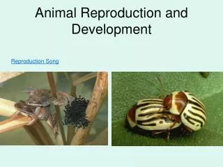
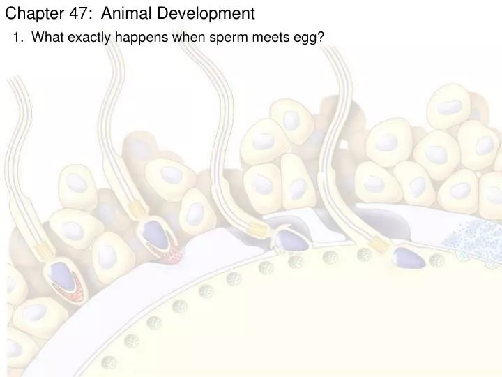
Chapter 47: Animal Development. What exactly happens when sperm meets egg?. 1. Acrosomal reaction. Hydrolytic enzymes released from the acrosome make a hole in the jelly coat, while growing actin filaments form the acrosomal process. This structure protrudes

E N D
Chapter 47: Animal Development • What exactly happens when sperm meets egg?
1 Acrosomal reaction. Hydrolytic enzymes released from the acrosome make a hole in the jelly coat, while growing actin filaments form the acrosomal process. This structure protrudes from the sperm head and penetrates the jelly coat, binding to receptors in the egg cell membrane that extend through the vitelline layer. 2 Contact and fusion of sperm and egg membranes. A hole is made in the vitelline layer, allowing contact and fusion of the gamete plasma membranes. The membrane becomes depolarized, resulting in the fast block to polyspermy. 3 5 4 Cortical reaction. Fusion of the gamete membranes triggers an increase of Ca2+ in the egg’s cytosol, causing cortical granules in the egg to fuse with the plasma membrane and discharge their contents. This leads to swelling of the perivitelline space, hardening of the vitelline layer, and clipping of sperm-binding receptors. The resulting fertilization envelope is the slow block to polyspermy. Contact. The sperm cell contacts the egg’s jelly coat, triggering exocytosis from the sperm’s acrosome. Entry of sperm nucleus. Sperm plasma membrane Sperm nucleus Acrosomal process Basal body (centriole) Fertilization envelope Sperm head Fused plasma membranes Cortical granule Actin Perivitelline space Hydrolytic enzymes Acrosome Cortical granule membrane Vitelline layer Jelly coat Egg plasma membrane Sperm-binding receptors EGG CYTOPLASM Figure 47.3 The acrosomal and cortical reactions during sea urchin fertilization
Chapter 47: Animal Development • What exactly happens when sperm meets egg? • Contact – acrosome releases hydrolytic enzymes • Acrosomal rxn – enzymes digest jelly coat while actin extends • - acrosomal process attaches to sperm binding receptors • Membrane fusion (sperm & egg) – causes depolarization as Ca+2 released • - aka fast block to polyspermy • - aka activation of the egg begins • Sperm nucleus enters egg • Cortical rxn – cortical granules from egg fuse with plasma membrane • Forms fertilization envelope aka slow block to polyspermy • What happens with activation of the egg? • Ca+2 released from ER • ↑ Cellular respiration & ↑protein synthesis (translation)
EXPERIMENT A fluorescent dye that glows when it binds free Ca2+ was injected into unfertilized sea urchin eggs. After sea urchin sperm were added, researchers observed the eggs in a fluorescence microscope. RESULTS 10 sec after fertilization 1 sec before fertilization 30 sec 20 sec Spreading wave of calcium ions Point of Sperm entry The release of Ca2+ from the endoplasmic reticulum into the cytosol at the site of sperm entry triggers the release of more and more Ca2+ in a wave that spreads to the other side of the cell. The entire process takes about 30 seconds. CONCLUSION Figure 47.4 What is the effect of sperm binding on Ca2+ distribution in the egg? 500 m
Binding of sperm to egg 1 2 Acrosomal reaction: plasma membrane depolarization (fast block to polyspermy) 3 4 6 Seconds 8 Increased intracellular calcium level 10 20 Cortical reaction begins (slow block to polyspermy) 30 40 50 Formation of fertilization envelope complete 1 2 Increased intracellular pH 3 4 5 Increased protein synthesis Minutes 10 Fusion of egg and sperm nuclei complete 20 30 Onset of DNA synthesis 40 60 First cell division 90 Figure 47.5 Timeline for the fertilization of sea urchin eggs
Chapter 47: Animal Development • What exactly happens when sperm meets egg? • What happens with activation of the egg? • What happens in mammals?
1 The sperm migrates through the coat of follicle cells and binds to receptor molecules in the zona pellucida of the egg. (Receptor molecules are not shown here.) This binding induces the acrosomal reaction, in which the sperm releases hydrolytic enzymes into the zona pellucida. 2 Breakdown of the zona pellucida by these enzymes allows the spermto reach the plasma membrane of the egg. Membrane proteins of the sperm bind to receptors on the egg membrane, and the two membranes fuse. 3 The nucleus and other components of the sperm cell enter the egg. 4 Follicle cell Sperm basal body Cortical granules Zone pellucida Sperm nucleus Enzymes released during the cortical reaction harden the zona pellucida, which now functions as a block to polyspermy. 5 Egg plasma membrane Acrosomal vesicle EGG CYTOPLASM Figure 47.6 Early events of fertilization in mammals
Chapter 47: Animal Development • What exactly happens when sperm meets egg? • What happens with activation of the egg? • What happens in mammals? • Contact – sperm migrates through follicle cells & binds to zona pellucida • Acrosomal rxn – acrosome releases hydrolytic enzymes digesting ZP • Sperm bind to sperm receptors on 2° oocyte & membranes fuse • Sperm nucleus enters egg • Cortical reaction hardens ZP as a block to polyspermy • What happens with during cleavage? • Cell division w/o cytokinesis • Creates blastomeres • Axes formed at first cleavage in amphibians
Figure 47.8 The body axes and their establishment in an amphibian Anterior Body axes. The three axes of the fully developed embryo, the tadpole, are shown above. (a) Right Dorsal Ventral Left Posterior Animal hemisphere Animal pole Point of sperm entry 1 The polarity of the egg determines the anterior-posterior axis before fertilization. Vegetal hemisphere Vegetal pole Point of sperm entry At fertilization, the pigmented cortex slides over the underlying cytoplasm toward the point of sperm entry. This rotation (red arrow) exposes a region of lighter-colored cytoplasm, the gray crescent, which is a marker of the dorsal side. 2 Future dorsal side of tadpole Gray crescent First cleavage The first cleavage division bisects the gray crescent. Once the anterior- posterior and dorsal-ventral axes are defined, so is the left-right axis. 3 (b) Establishing the axes. The polarity of the egg and cortical rotation are critical in setting up the body axes.
Zygote 0.25 mm 2-cell stage forming Eight-cell stage (viewed from the animal pole). The large amount of yolk displaces the third cleavage toward the animal pole, forming two tiers of cells. The four cells near the animal pole (closer, in this view) are smaller than the other four cells (SEM). 4-cell stage forming 8-cell stage 0.25 mm Blasto- coel Animal pole Blastula (at least 128 cells). As cleavage continues, a fluid-filled cavity, the blastocoel, forms within the embryo. Because of unequal cell division due to the large amount of yolk in the vegetal hemisphere, the blastocoel is located in the animal hemisphere, as shown in the cross section. The SEM shows the outside of a blastula with about 4,000 cells, looking at the animal pole. Blastula (cross section) Vegetal pole Figure 47.9 Cleavage in a frog embryo
Chapter 47: Animal Development • What exactly happens when sperm meets egg? • What happens with activation of the egg? • What happens in mammals? • What happens with during cleavage? • What is gastrulation? • Movement of blastula cells into the blastopore creating 2 cell (germ) layers • Ectoderm – outer layer • Endoderm – inner layer • Mesoderm – forms in between
CROSS SECTION SURFACE VIEW Gastrulation begins when a small indented crease, the dorsal lip of the blastopore, appears on one side of the blastula. The crease is formed by cells changing shape and pushing inward from the surface (invagination). Additional cells then roll inward over the dorsal lip (involution) and move into the interior, where they will form endoderm and mesoderm. Meanwhile, cells of the animal pole, the future ectoderm, change shape and begin spreading over the outer surface. Animal pole Blastocoel Dorsal lip of blastopore Dorsal lip of blastopore Blastula Vegetal pole Archenteron Blastocoel shrinking 3 2 The blastopore lip grows on both sides of the embryo, as more cells invaginate. When the sides of the lip meet, the blastopore forms a circle that becomes smaller as ectoderm spreads downward over the surface. Internally, continued involution expands the endoderm and mesoderm, and the archenteron begins to form; as a result, the blastocoel becomes smaller. Ectoderm Late in gastrulation, the endoderm-lined archenteron has completely replaced the blastocoel and the three germ layers are in place. The circular blastopore surrounds a plug of yolk-filled cells. Blastocoel remnant Mesoderm Endoderm Key Future ectoderm 1 Future mesoderm Yolk plug Yolk plug Gastrula Future endoderm Figure 47.12 Gastrulation in a frog embryo
Chapter 47: Animal Development • What exactly happens when sperm meets egg? • What happens with activation of the egg? • What happens in mammals? • What happens with during cleavage? • What is gastrulation? • What is organogenesis? • Creation of organs • Involves folds, splits & clustering of cells • 1st organs are neural tube & notocord
Chapter 47: Animal Development Students Correlations available now – sorry for the delay Learning Log – later today AP checks?? – March 9 deadline Has anyone not taken the Biology EOC? Transfers, movers,
Neural folds Eye Somites Tail bud Neural fold Neural plate SEM Neural tube 1 mm LM 1 mm Notochord Neural crest Neural fold Neural plate Neural crest Coelom Somite Notochord Ectoderm Mesoderm Outer layer of ectoderm Archenteron (digestive cavity) Endoderm Neural crest Archenteron (c) Somites. The drawing shows an embryo after completion of the neural tube. By this time, the lateral mesoderm has begun to separate into the two tissue layers that line the coelom; the somites, formed from mesoderm, flank the notochord. In the scanning electron micrograph, a side view of a whole embryo at the tail-bud stage, part of the ectoderm has been removed, revealing the somites, which will give rise to segmental structures such as vertebrae and skeletal muscle. (a) Neural plate formation. By the time shown here, the notochord has developed from dorsal mesoderm, and the dorsal ectoderm has thickened, forming the neural plate, in response to signals from the notochord. The neural folds are the two ridges that form the lateral edges of the neural plate. These are visible in the light micrograph of a whole embryo. Neural tube (b) Formation of the neural tube. Infolding and pinching off of the neural plate generates the neural tube. Note the neural crest cells, which will migrate and give rise to numerous structures. Figure 47.14 Early organogenesis in a frog embryo
ECTODERM MESODERM ENDODERM • Epidermis of skin and itsderivatives (including sweatglands, hair follicles) • Epithelial lining of mouthand rectum • Sense receptors inepidermis • Cornea and lens of eye • Nervous system • Adrenal medulla • Tooth enamel • Epithelium or pineal andpituitary glands • Notochord • Skeletal system • Muscular system • Muscular layer of stomach, intestine, etc. • Excretory system • Circulatory and lymphaticsystems • Reproductive system(except germ cells) • Dermis of skin • Lining of body cavity • Adrenal cortex • Epithelial lining ofdigestive tract • Epithelial lining ofrespiratory system • Lining of urethra, urinarybladder, and reproductivesystem • Liver • Pancreas • Thymus • Thyroid and parathyroidglands Figure 47.16 Adult derivatives of the three embryonic germ layers in vertebrates
Chapter 47: Animal Development • What exactly happens when sperm meets egg? • What happens with activation of the egg? • What happens in mammals? • What happens with during cleavage? • What is gastrulation? • What is organogenesis? • What are the 4 extra-embryonic membranes in the amniotic egg? • Amnion • Allantois • Chorion • Yolk sac
Allantois. The allantois functions as a disposal sac for certain metabolic wastes produced by the embryo. The membrane of the allantois also functions with the chorion as a respiratory organ. Amnion. The amnion protects the embryo in a fluid-filled cavity that prevents dehydration and cushions mechanical shock. Embryo Albumen Amniotic cavity with amniotic fluid Yolk (nutrients) Shell Chorion. The chorion and the membrane of the allantois exchange gases between the embryo and the surrounding air. Oxygen and carbon dioxide diffuse freely across the egg’s shell. Yolk sac. The yolk sac expands over the yolk, a stockpile of nutrients stored in the egg. Blood vessels in the yolk sac membrane transport nutrients from the yolk into the embryo. Other nutrients are stored in the albumen (the ”egg white”). Figure 47.17 Extraembryonic membranes in birds and other reptiles
Chapter 47: Animal Development • What exactly happens when sperm meets egg? • What happens with activation of the egg? • What happens in mammals? • What happens with during cleavage? • What is gastrulation? • What is organogenesis? • What are the 4 extra-embryonic membranes in the amniotic egg? • How does mammalian development occur? • Slow cleavage • 1st division – 36 hrs • 2nd – 60 hrs • 3rd – 72 hrs
Endometrium (uterine lining) Inner cell mass Trophoblast Blastocoel Blastocyst reaches uterus. 4 2 1 3 Expanding region of trophoblast Maternal blood vessel Epiblast Hypoblast Trophoblast Blastocyst implants. Expanding region of trophoblast Amniotic cavity Amnion Epiblast Hypoblast Chorion (from trophoblast) Extraembryonic membranes start to form and gastrulation begins. Extraembryonic mesoderm cells (from epiblast) Yolk sac (from hypoblast) Amnion Allantois Chorion Ectoderm Mesoderm Endoderm Yolk sac Gastrulation has produced a three- layered embryo with four extraembryonic membranes. Extraembryonic mesoderm Figure 47.18 Four stages in early embryonic development of a human
Chapter 47: Animal Development • What exactly happens when sperm meets egg? • What happens with activation of the egg? • What happens in mammals? • What happens with during cleavage? • What is gastrulation? • What is organogenesis? • What are the 4 extra-embryonic membranes in the amniotic egg? • How does mammalian development occur? • What three things influence cell fate? • Cytoplasmic determinants – mRNA & proteins in egg cytoplasm • Induction – cellular peer pressure • Cleavage pattern – divides cytoplasmic determinants
Unfertilized egg cell Sperm Molecules of another cyto- plasmic deter- minant Molecules of a a cytoplasmic determinant Fertilization Zygote (fertilized egg) Mitotic cell division Two-celled embryo (a) Cytoplasmic determinants in the egg. The unfertilized egg cell has molecules in its cytoplasm, encoded by the mother’s genes, that influence development. Many of these cytoplasmic determinants, like the two shown here, are unevenly distributed in the egg. After fertilization and mitotic division, the cell nuclei of the embryo are exposed to different sets of cytoplasmic determinants and, as a result, express different genes. Figure 21.11 Sources of developmental information for the early embryo Nucleus
Early embryo (32 cells) Signal transduction pathway NUCLEUS Signal receptor Signal molecule (inducer) Induction by nearby cells. The cells at the bottom of the early embryo depicted here are releasing chemicals that signal nearby cells to change their gene expression. (b) Figure 21.11b Cellular peer pressure
CONCLUSION EXPERIMENT RESULTS 1 2 The totipotency of the two blastomeres normally formed during the first cleavage division depends on cytoplasmic determinants localized in the gray crescent. Figure 47.24 How does distribution of the gray crescent at the first cleavage affect the potency of the two daughter cells? Left (control): Fertilized salamander eggs were allowed to divide normally, resulting in the gray crescent being evenly divided between the two blastomeres. Right (experimental): Fertilized eggs were constricted by a thread so that the first cleavage plane restricted the gray crescent to one blastomere. Gray crescent Gray crescent The two blastomeres were then separated and allowed to develop. Belly piece Normal Normal Blastomeres that receive half or all of the gray crescent develop into normal embryos, but a blastomere that receives none of the gray crescent gives rise to an abnormal embryo without dorsal structures. Spemann called it a “belly piece.”
Spemann and Mangold transplanted a piece of the dorsal lip of a pigmented newt gastrula to the ventral side of the early gastrula of a nonpigmented newt. CONCLUSION EXPERIMENT RESULTS Pigmented gastrula (donor embryo) Dorsal lip of blastopore Nonpigmented gastrula (recipient embryo) During subsequent development, the recipient embryo formed a second notochord and neural tube in the region of the transplant, and eventually most of a second embryo. Examination of the interior of the double embryo revealed that the secondary structures were formed in part from host tissue. Primary embryo Primary structures: Secondary (induced) embryo Secondary structures: Neural tube Notochord Notochord (pigmented cells) Neural tube (mostly nonpigmented cells) The transplanted dorsal lip was able to induce cells in a different region of the recipient to form structures different from their normal fate. In effect, the dorsal lip “organized” the later development of an entire embryo. Figure 47.25 Can the dorsal lip of the blastopore induce cells in another part of the amphibian embryo to change their developmental fate?
Chapter 47: Animal Development • What exactly happens when sperm meets egg? • What happens with activation of the egg? • What happens in mammals? • What happens with during cleavage? • What is gastrulation? • What is organogenesis? • What are the 4 extra-embryonic membranes in the amniotic egg? • How does mammalian development occur? • What three things influence cell fate? • How are organisms formed from the fertilized egg? • Cell division/cleavage • Morphogenesis – process of giving shape to an organism • Cell differentiation – process by which cells become specialized
(a) Animal development. Most animals go through some variation of the blastula and gastrula stages. The blastula is a sphere of cells surrounding a fluid-filled cavity. The gastrula forms when a region of the blastula folds inward, creating a tube—a rudimentary gut. Once the animal is mature, differentiation occurs in only a limited way—for the replacement of damaged or lost cells. Gut Cell movement Zygote (fertilized egg) Eight cells Blastula (cross section) Gastrula (cross section) Adult animal (sea star) Cell division Morphogenesis Observable cell differentiation (b) Plant development. In plants with seeds, a complete embryo develops within the seed. Morphogenesis, which involves cell division and cell wall expansion rather than cell or tissue movement, occurs throughout the plant’s lifetime. Apical meristems (purple) continuously arise and develop into the various plant organs as the plant grows to an indeterminate size. Seed leaves Shoot apical meristem Root apical meristem Zygote (fertilized egg) Two cells Embryo inside seed Plant Figure 21.4 Some key stages of development in animals and plants
Chapter 47: Animal Development • What exactly happens when sperm meets egg? • What happens with activation of the egg? • What happens in mammals? • What happens with during cleavage? • What is gastrulation? • What is organogenesis? • What are the 4 extra-embryonic membranes in the amniotic egg? • How does mammalian development occur? • What three things influence cell fate? • How are organisms formed from the fertilized egg? • Can cells de-differentiate? • Plant cuttings • Animal cells????
Researchers enucleated frog egg cells by exposing them to ultraviolet light, which destroyed the nucleus. Nuclei from cells of embryos up to the tadpole stage were transplanted into the enucleated egg cells. EXPERIMENT Frog tadpole Frog embryo Frog egg cell Fully differ- entiated (intestinal) cell Less differ- entiated cell Donor nucleus trans- planted Enucleated egg cell Donor nucleus trans- planted <2% develop into tadpoles Most develop into tadpoles Figure 21.6 Can the nucleus from a differentiated animal cell direct development of an organism?
Chapter 47: Animal Development • What exactly happens when sperm meets egg? • What happens with activation of the egg? • What happens in mammals? • What happens with during cleavage? • What is gastrulation? • What is organogenesis? • What are the 4 extra-embryonic membranes in the amniotic egg? • How does mammalian development occur? • What three things influence cell fate? • How are organisms formed from the fertilized egg? • Can cells de-differentiate? • How was Dolly cloned? • - Nuclear transplantation
Egg cell donor Mammary cell donor APPLICATION This method is used to produce cloned animals whose nuclear genes are identical to the donor animal supplying the nucleus. 1 2 Egg cell from ovary Nucleus removed Nucleus removed Cells fused Cultured mammary cells are semistarved, arresting the cell cycle and causing dedifferentiation 3 TECHNIQUE Shown here is the procedure used to produce Dolly, the first reported case of a mammal cloned using the nucleus of a differentiated cell. Nucleus from mammary cell Grown in culture 4 RESULTS The cloned animal is identical in appearance and genetic makeup to the donor animal supplying the nucleus, but differs from the egg cell donor and surrogate mother. Early embryo Implanted in uterus of a third sheep 5 Surrogate mother Embryonic development 6 Lamb (“Dolly”) genetically identical to mammary cell donor Fig. 21.7 Reproductive Cloning of a Mammal by Nuclear Transplantation
Embryonic stem cells Adult stem cells Early human embryo at blastocyst stage (mammalian equiva- lent of blastula) From bone marrow in this example Totipotent cells Pluripotent cells Cultured stem cells Different culture conditions Liver cells Blood cells Nerve cells Different types of differentiated cells Figure 21.9 Working with stem cells
Chapter 47: Animal Development • What exactly happens when sperm meets egg? • What happens with activation of the egg? • What happens in mammals? • What happens with during cleavage? • What is gastrulation? • What is organogenesis? • What are the 4 extra-embryonic membranes in the amniotic egg? • How does mammalian development occur? • What three things influence cell fate? • How are organisms formed from the fertilized egg? • Can cells de-differentiate? • How was Dolly cloned? • When is a cell determined (fated)? • - Muscle cells – MyoD transcription factor – turns on all muscle genes
Nucleus Master control gene myoD Other muscle-specific genes DNA OFF OFF Embryonicprecursor cell Figure 21.10 Determination and differentiation of muscle cells
1 Nucleus Master control gene myoD Other muscle-specific genes DNA OFF OFF Embryonicprecursor cell Determination. Signals from othercells lead to activation of a masterregulatory gene called myoD, andthe cell makes MyoD protein, atranscription factor. The cell, nowcalled a myoblast, is irreversiblycommitted to becoming a skeletalmuscle cell. OFF mRNA MyoD protein(transcriptionfactor) Myoblast (determined) Figure 21.10 Determination and differentiation of muscle cells
1 2 Nucleus Master control gene myoD Other muscle-specific genes DNA OFF OFF Embryonicprecursor cell Determination. Signals from othercells lead to activation of a masterregulatory gene called myoD, andthe cell makes MyoD protein, atranscription factor. The cell, nowcalled a myoblast, is irreversiblycommitted to becoming a skeletalmuscle cell. OFF mRNA MyoD protein(transcriptionfactor) Myoblast (determined) Differentiation. MyoD protein stimulatesthe myoD gene further, and activatesgenes encoding other muscle-specifictranscription factors, which in turn activate genes for muscle proteins. MyoD also turns on genes that block the cell cycle, thus stopping cell division. The nondividing myoblasts fuse to become mature multinucleate muscle cells, alsocalled muscle fibers. mRNA mRNA mRNA mRNA Myosin, othermuscle proteins,and cell-cycleblocking proteins MyoD Anothertranscriptionfactor Muscle cell(fully differentiated) Figure 21.10 Determination and differentiation of muscle cells
Chapter 47: Animal Development • What exactly happens when sperm meets egg? • What happens with activation of the egg? • What happens in mammals? • What happens with during cleavage? • What is gastrulation? • What is organogenesis? • What are the 4 extra-embryonic membranes in the amniotic egg? • How does mammalian development occur? • What three things influence cell fate? • How are organisms formed from the fertilized egg? • Can cells de-differentiate? • How was Dolly cloned? • When is a cell determined (fated)? • What is apoptosis? • - Programmed cell death – cell suicide
Ced-9 protein (active) inhibits Ced-4 activity Mitochondrion Ced-4 Ced-3 Death signal receptor Inactive proteins Cell forms blebs (a) No death signal Ced-9 (inactive) Death signal Active Ced-3 Active Ced-4 Other proteases Nucleases Activation cascade (b) Death signal Figure 21.18 Molecular basis of apoptosis in C. elegans
Chapter 47: Animal Development • What exactly happens when sperm meets egg? • What happens with activation of the egg? • What happens in mammals? • What happens with during cleavage? • What is gastrulation? • What is organogenesis? • What are the 4 extra-embryonic membranes in the amniotic egg? • How does mammalian development occur? • What three things influence cell fate? • How are organisms formed from the fertilized egg? • Can cells de-differentiate? • How was Dolly cloned? • When is a cell determined (fated)? • What is apoptosis? • What are some model organisms for studying development?
DROSOPHILA MELANOGASTER (FRUIT FLY) CAENORHABDITIS ELEGANS (NEMATODE) Figure 21.2 Model Organisms for Genetic Studies of Development Drosophila - small, easy & cheap to culture - 2 week generation time - 4 chromosomes - LARGE literature of info C elegans - easy to culture - transparent body with few cell types - zygote to mature adult in 3 days 0.25 mm
ARABIDOPSIS THAMANA (COMMON WALL CRESS) MUS MUSCULUS (MOUSE) DANIO RERIO (ZEBRAFISH) Mouse - vertebrate - LARGE literature - transgenics & knock-outs