Download
1 / 20
280 likes | 622 Views
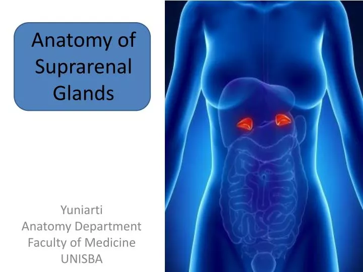
Anatomy of Suprarenal Glands. Yuniarti Anatomy Department Faculty of Medicine UNISBA. Location of Suprarenal Glands Located between the superomedial aspects of the kidneys and the diaphragm.

E N D
Anatomy of Suprarenal Glands Yuniarti Anatomy Department Faculty of Medicine UNISBA
Location • of Suprarenal Glands • Located between the superomedial aspects of the kidneys and the diaphragm
The pyramidal right gland is more apical (situated over the superior pole) relative to the right kidney, lies anterolateral to the right of the diaphragm, and makes contact with the IVC anteromedially and the liver anterolaterally
The crescent-shaped left gland is medial to the superior half of the left kidney and is related to the spleen, stomach, pancreas and the left of the diaphragm
Suprarenal glands surrounded by connective tissue containing • considerable perinephric fat • Suprarenal glands are enclosed by renal fascia by which they are attached to • the crura of the diaphragm • Each gland has a hilum, where the veins and lymphatic vessels exit the gland; • whereas arteries and nerve enter the glands at multiple sites • They are separated from the kidneys by a thin septum(part of the renal fascia)
The medial borders of the suprarenal glands are 4-5 cm apart. In this area, • from right to left, are the IVC, right of the diaphragm, celiac ganglion, • celiac trunk, SMA and the left of the diaphragm.
Each suprarenal gland has two parts : • ~ Suprarenal cortex • ~ Suprarenal medulla
SUPRARENAL CORTEX • Derives from mesoderm
Glucocorticoid • include cortisol (hydrocortisone),corticosterone, and cortisone • Function : Protein breakdown, glucose formation, lipolysis, resistance to stress, anti-inflammatory effects, depression of immune responses
Androgen • The major androgen secreted by the adrenal gland is dehydroepiandrosterone (DHEA) • Function : promote libido (sex drive) and are convertedinto estrogens (feminizing sex steroids) by other body tissues; after menopause, when ovarian secretion of estrogens ceasesall female estrogens come from conversion of adrenal androgens; stimulate growth of axillary andpubic hair in boys and girls and contribute to the prepubertalgrowth spurt.
SUPRARENAL MEDULLA • Derive from neural crest cells associated • with the sympathetic nervous system • The cromaffin cells secrete catecholamines / epinephrine and norepinephrine (NE), also • called adrenaline and noradrenaline, respectively. • Unlike the hormones of the adrenal cortex, the hormonesof the adrenal medulla are not essential for life sincethey only intensify sympathetic responses in other parts of the body.
Action of catecholamine : • Increased heart rate • Increased force of cardiac muscle contraction • Elevated blood pressure • Increased breathing rate • Decreased activity in the digestive system
Vascularisation of suprarenal glands Suprarenal glands arteries : ~ Superior suprarenal arteries ~ Middle suprarenal arteries ~ Inferior suprarenal arteries
Venous drainage : ~ Right suprarenal vein drains into the IVC ~ Left suprarenal vein drains into the left renal vein
Lymphatic Suprarenal Glands The lymph passes to the right & left lumbar lymph nodes
Nerves of the suprarenal glands • Celiac plexus • Abdominopelvic (greater, lesser and least) splanchnic nerve
Myelinated presynaptic sympathetic fibers mainly derived from lateral horn of gray matter of the spinal cord segments T10-L1 -- transverse both the paravertebral and the prevertebral ganglia, without synapse, to be distributed to the chromaffin cells in the suprarenal medulla.