Download
1 / 23
230 likes | 381 Views
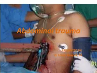
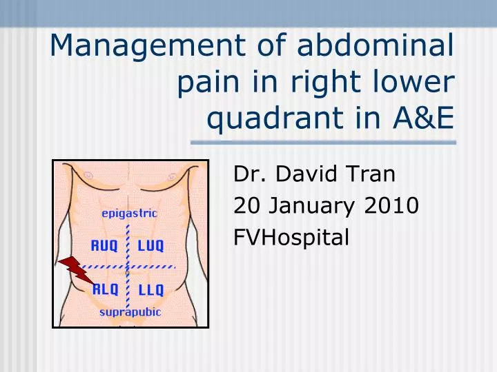
Management of abdominal pain in right lower quadrant in A&E. Dr. David Tran 20 January 2010 FVHospital. Short case report:. Man 76 years old, abdominal pain for 48h. Physical exam: pain at the right flanck, right hypochondre and right lower quadrant (tenderness). WBC 13.800, CRP 184

E N D
Management of abdominal pain in right lower quadrant in A&E Dr. David Tran 20 January 2010 FVHospital
Short case report: • Man 76 years old, abdominal pain for 48h. • Physical exam: pain at the right flanck, right hypochondre and right lower quadrant (tenderness). • WBC 13.800, CRP 184 • ASP Xray: normal,no hydro-aeric level
Abdominal Ultrasound 2/ A l'étage pelvien: • L'examen a été réalisé par voie sus-pubienne. • Vessie anéchogène, à parois fines. • Les coupes réalisées au niveau du pelvis montrent une prostate de volume normal, de contours réguliers et nets. Sa structure échographique est homogène. • FID sans particularite Au total: • Examen normal.
Abdominal CT scanner • Présence d'une infiltration graisseuse en dessous du caecum associée aux bulles d'air extradigestives. • Présence de diverticules sigmoidiens. • Pas d'épanchement liquidien péritonéal. • Pas de pneumoperitoine libre. • Le reste de l'examen est sans particularite. Conclusion: • Péritonite localisée de la fosse iliaque droite, d'origine d'une rupture soit appendiculaire, soit diverticulaire. • Diverticules sigmoidiens.
Suspected appendicitis • Historical management: early laparotomy to avoid risk of appendix perforation. • In 20% of patients who undergo exploratory laparotomy, appendix = normal. • Elderly patients and female > the error rate is about 40%
Strategy if pain at the right lower quadrant • Medical history & physical examination is the cornerstone in evaluation • 3 Common signs of appendicitis may support the diagnosis: • Pain at the right lower quadrant (RLQ) • Guarding at palpation RLQ > Abdominal rigidity • Migration of pain from periumbilical region
Anatomic basis of Psoas sign • Inflate appendix is in the retroperitoneal location in contact with the psoas muscle • The appendix is stretched by the extension of the psoas muscle.
Definition of Psoas sign • Pain on passive extension of the right thigh, patient lies on left side. Examiner extends patient’s right thigh while applying counter resistance to the right hip.
Definition of Obturator sign • Pain on passive internal rotation of the flexed thigh. Examiner moves lower leg laterally while applying resistance to the lateral side of the knee.
Anatomic basis of the Obturator sign • Inflamed appendix in the pelvis is in contact with the obturator internus muscle • The Obturator is stretched by the maneuver
Signs of peritoneal inflammation • Involuntary rigidity or spasm at the abdominal muscles • Rebound tenderness (It hurts more when you release pressure) • Coughing increase the abdominal pain
Most common misdiagnosis • Gastroenteritis • Urinary tract infection • Renal colic • Rupture ovarian follicle • Ectopic pregnancy Young women
Laboratory testing • WBC & CRP are useful to confirm inflammatory syndrome (but poor specificity for appendicitis) • Urinalysis must be done (can show blood or leucocytes) • Beta HCG must be search for all women in reproductive age (ectopic pregnancy?)
Conventional radiology • Law sensitivity and specificity for the diagnosis of acute appendicitis… • Appendicolith is very rare • ASP shouldn’t be ordered if suspected appendicitis, except if there is an occlusive syndrome.
Ultrasound • Ultrasound has Se 75-90 and Sp 86-100 • May identify alternative diagnosis like pyosalpynx or ovarian cyst. • Appendicitis may be rule out if the appendix is normal on ultrasound. • The failure to see the appendix limits the usefulness of ultrasound
Criteria for ultrasound diagnosis of appendicitis are : 1. non-compressible aperistaltic sausage appendix with wall thickening. Ultrasound findings in non-perforated appendicitis include a muscular wall thickness greater than 2 mm, an appendicial diameter (outer wall to outer wall) greater than 7 mm that does not compress, a "target" sign (bull's-eye appearance) of abnormally thickened bowel wall layers when viewed in the short axis, and sometimes distension or obstruction of the appendicial lumen accompanied by increased echogenicity "oedema" surrounding the appendix. Findings may also include Doppler in the wall of the appendix, indicating increased appendicial perfusion. 2. démonstration of an appendicolith, which is seen as an echogenic focus within the appendix lumen with shadowing.3. Further signs include fluid around the appendix, an inflammatory bowel mass and the formation of abscess. Criteria for diagnosis of appendicitis in ultrasound • 1. non-compressible sausage appendix with wall thickening. Ultrasound findings in non-perforated appendicitis include a muscular wall thickness greater than 2 mm, an appendicial diameter greater than 7 mm that does not compress, abnormally thickened bowel wall when viewed in the short axis, and sometimes distension or obstruction of the appendicial lumen accompanied by increased echogenicity "oedema" surrounding the appendix. • 2.démonstration of an appendicolith, which is seen as an echogenic focus within the appendix lumen with shadowing. • 3.Further signs include fluid around the appendix, an inflammatory bowel mass and the formation of abscess.
Computed Tomography • Se 90-100% • Sp 91-99% • Distended appendix • Thickened appendiceal wall • Preiappendiceal inflammation
CT scanner or ultrasound ? • Greater Se of CT (96% versus 76%) • Higher negative predictive value for CT (95% versus 76%) • Alternative diagnosis more often with CT. • Appendix often not seen in ultrasound… • Superiority of CT in diagnosis of appendicitis
Conclusion • Physical exam remains the corner stone of the diagnosis of appendicitis • Conventional Xray is useless for diagnosis (except rare appendicolith) • WBC and CRP help the diagnosis but have poor Se & Sp. • If equivocal presentation, CT scanner is better than ultrasound