Download
1 / 1

E N D
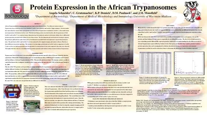
DISCUSSION • The first key results showed that their were proteins detectable on one of the gels that did not occur on the other gel. This is interesting because one would not expect to find very many proteins that are different in trypanosomes clonally related like LouTat 1 and LouTat 1A, because most proteins probably represent housekeeping gene expression within the cells. • The fact that protein differences exist leads to the possibility of determining amino acid sequence, the specific protein, and then finding out which gene is responsible for encoding that protein. The discovery of which gene(s) is (are) controlling specific differentially expressed proteins could lead to an understanding how the different strains of African Trypanosomes express differing virulence levels. Once genes are identified that regulate potential virulence protein expression, they can be manipulated by deletion, knockdown or even overexpression; such mutated trypanosomes can then be used for infection of mice to determine the phenotypic levels of virulence expressed. Protein Expression in the African TrypanosomesAngela Schneider1, C. Grutzmacher1, K.P. Demick1, D.M. Paulnock2, and J.M. Mansfield11Department of Bacteriology, 2Department of Medical Microbiology and Immunology University of Wisconsin-Madison INTRODUCTION Trypanosoma brucei rhodesiense is an extracellular protozoan parasite spread by bite of Glossina (tsetse fly) that has caused tens of thousands of human deaths each year in sub-Saharan Africa (WHO Expert Committee on the Control and Surveillance of African Trypanosomiasis, 1998). This parasitic infection is fatal. The immune system is unable to control the disease due to antigenic variation in the variant surface glycoprotein coat (VSG) of the parasite (Dubois et al., 2005). The role of the VSG protein coat in providing an escape mechanism for trypanosomes generated a question as to whether different virulence levels displayed by African trypanosomes were linked to VSG protein coat expression. It was found that neither the VSG genes nor the protein coat made a difference in how virulent the strains were (Inverso, 2010). My projecthasaddressed the hypothesis that different genes are being turned on inside the cell at different times and therefore are able to express different proteins in the different virulent strains LouTat 1 and LouTat 1A. My experiments analyzed the level of expression of proteins between the trypanosome strains; since both strains express the same VSG coat, only non-coat proteins that are different were detected. Figure 3: 2D gel representation of total cell protein extracts from Lou Tat 1. The red outlines represent spots that were expressed only on the gel from LouTat 1 and not on the gel from LouTat1A. The green outlines represent spots on the gel from LouTat 1 that show a two-fold increase in expression levels from the spots on the gel for LouTat 1A . Figure 4: 2D gel representation of total cell protein extracts from LouTat1A. The red outlines represent spots that were only expressed on the gel from LouTat1A but not on the gel from LouTat 1. The green outlined spots represent spots on the gel for LouTat 1A that show a two-fold increase in expression levels from the spots on the gel for LouTat 1. ABSTRACT African Trypanosomiasis is a fatal parasitic infection found in Sub-Saharan Africa. Two different virulent strains of Trypanosoma bruceirhodesiensehave been identified,LouTat 1 (low virluence) and LouTat 1A (high virulence). Previous studies have shown that the variant surface glycoprotein coat of the African Trypanosome is unrelated to virulence, so the model system uses trypanosomes with identical surface coats. With this knowledge protein extraction from the cells of trypanosomes of both LouTat 1 and LouTat 1A were analyzed using 2 dimensional gel electrophoresis analysis to determine whether there is differential protein expression associated with virulence levels of these strains. The first dimension gels used isoelectric focusing to separate the proteins by pH/isoelectric point whereas the second dimension used SDS PAGE electrophoresis to separate the proteins by mass. The gels from LouTat 1 and LouTat 1A were then analyzed by the computer imaging and analysis software, Phoretix, to detect differences in protein expression. The existence of certain unique protein spots on LouTat 1 gels in comparison to LouTat 1A gels as well as two fold increases/decreases in expression of certain proteins of one strain compared to the other were observed. This implies that there may be a correlation between protein expression and virulence in these strains of African Trypanosomes. Figure 5: Graphical representation of variation in protein spot expression in LouTat1 and LouTat 1A. This figure is derived from a bar graph representation of the variation in expression of each spot on the gel. The reference gel for this figure was LouTat 1. Figure 6: Mass spectrometry analysis of a differentially expressed protein. This figure represents the spectral analysis of a protein spot labeled number one identified on the gels for LouTat 1 vsLouTat 1A. The results of this analysis found that the protein identified was Beta Tubulin. • PHORETIX RESULTS • The gels revealed protein differences between the two strains, LouTat 1 and LouTat 1A , but there were primarily common proteins detected. • The analysis of the gels also showed that there were some protein spots that were expressed in one strain of the organism but were not expressed in the other strain. • The analysis also showed that there were multiple proteins detectable on the gel for LouTat 1 that occurred at either at a two-fold increase or decrease compared to corresponding spots on the LouTat 1A gel or vice versa. • MASS SPECTROMETRY PRELIMINARY RESULTS • Preliminary mass spectrometry data shows that Beta Tublin is a major protein that shows increased expression in LouTat 1 versus LouTat 1A. • Another protein identified as showing increased expression in LouTat 1 versus LouTat 1A is the variant surface glycoprotein. Methods Mice were infected with either the LouTat 1 or the LouTat 1A strains of African Trypanosomes. After 5 days the mice were sacrificed, bled out, the trypanosomes purified from blood and the proteins were extracted. The first dimension of the analysis used an isoelectric focusing machine to separate the proteins using pH gradients. The second dimension of the project used gel electrophoresis to separate proteins by mass. Once the gels were run they were placed in a fixing solution and then a CoomassieBlue stain was used to detect proteins after rinsing and destaining. The analysis of stained gels was done by using the computer imaging and 2D gel analysis program Phoretix, which allows the user to compare gels to each other to identify differential protein spot expression. Figure 1: Hoefer isoelectric focusing apparatus. The figure to the left is an IPGphormachine; this is used for the first dimension electrophoretic separation of trypanosome proteins based on isoelectric points. ACKNOWLEDGEMENTS Angela Schneider would like to acknowledge the help of Doctor Mansfield, Karen Demick, Cathy Grutzmacher and Gina Hedberg, members of the Mansfield/Paulnock lab in the Department of Bacteriology and the Department of Medical Microbiology and Immunology at the University of Wisconsin- Madison, for their help as well as their insightful guidance in regards to this experiment. REFERENCES Dubois, M.E., Demick, K.P. and Mansfield, J.M. (2005). Trypanosomes expressing a mosaic variant surface glycoprotein coat escape early detection by the immune system. Infect Immun 73, 2690-2697. Inverso, J.A., T.S. Uphoff, et al. (2010) Biological variation among African trypanosomes. I. Virulence regulation is not linked to the variant surface glycoprotein (VSG) or the VSG gene telomeric expression site. WHO Expert Committee on the Control and Surveillance of African Trypanosomiasis (1998) Control and surveillance of African trypanosomiasis : report of a WHO Expert Committee Geneva, World Health Organization, pp. vii, 113 p. Wren B. Deciphering tsetse’s secret partner [Online]. <http://jcs.biologists.org/content/vol115/issue12/cover.shtml>. [November 15, 2009]. Figure 2: SDS PAGE Gel Electrophoresis. The figure to the right is a gel electrophoresis apparatus used to separate proteins based on molecular mass.