Download
1 / 2
20 likes | 163 Views
Supplementary Fig. 1. 76N.TERT.Ecd. 76N.TERT.V. a. MEF. Ecd +/+. c. MEF.Ecd -/-. IgG. IgG. Ecd antibody. Ecd antibody. MEF mEcd +/+. 76N.TERT.V. 76N.TERT.Ecd. b. d. MEF mEcd -/-. hEcd. mEcd. β- actin. β- actin.

E N D
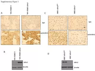
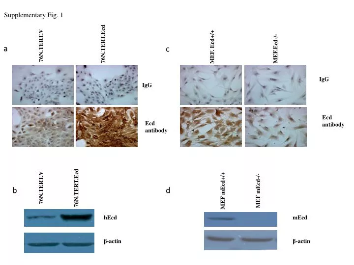
Supplementary Fig. 1 76N.TERT.Ecd 76N.TERT.V a MEF. Ecd+/+ c MEF.Ecd-/- IgG IgG Ecd antibody Ecd antibody MEF mEcd+/+ 76N.TERT.V 76N.TERT.Ecd b d MEF mEcd-/- hEcd mEcd β-actin β-actin
Supplementary Fig.1Characterization of Ecd antibody specificity for IHC staining. (A). 76NTERT cells expressing either vector (76NTERT.V) or Ecd (76NTERT.Ecd) were analyzed by IHC staining using either mouse IgG (as control) or anti-Ecd antibodies (B) Western blot analysis of cells used in Fig. A (C). IHC staining of mouse embryonic fibroblasts expressing wild type Ecd (Ecd+/+) or cre-adenovirus infected MEFs where Ecd was deleted (Ecd-/-). Mouse IgG was used as negative control. (D) Western blotting of cells used in Figure C.