Download
1 / 17
170 likes | 309 Views
Chlamydiae. Physiology and strucutre 1. Elementary body (EB) - Extracellular phase, small metabolically inactive, disulfide linkages in outer cell wall. 2. Reticulate body (RB) - Intracellular, replicative phase, metabolically active, osmotically fragile.

E N D
Chlamydiae Physiology and strucutre 1. Elementary body (EB) - Extracellular phase, small metabolically inactive, disulfide linkages in outer cell wall. 2. Reticulate body (RB) - Intracellular, replicative phase, metabolically active, osmotically fragile. 3. EB posseses genus and strain specific antigens in the outer coat. 4. Chlamydiae are non-motile and non-fimbriate.
Sequence of infectivity 1. Attachment by EB 2. Induced phagocytosis 3. EB RB conversion 4. Replication in phagosome 5. RB------- EB conversion and release
Entry and attachment 1. EBs may enter non-phagocytic cells 2. Purified cell-free, cell walls of EB can be internalized 3. EB surfaces are both hydrophobic and negatively charged. 4. EB coat is rich in outer membrane proteins that are extensively cross-linked by S-S bonds. 5. Rate of infection is proportional to the number of EBs adhering. 6. Specific receptor/ligand interaction??
Entry and attachment contd. 7. Protease treatment of EBs does not appear to decrease attachment, periodate treatment does. 8. Polyclonal antibodies to outer membrane proteins will also inhibit attachment.
Internalization 1. Energy required by host cell but not EB, therefore ingestion blocked by cyanide, etc. 2. Host cell protein synthesis is not required. 3. 2-3 hours post-attachment EBs appear within membrane-bound vacuoles. 4. Using polarized cells, microfilament formation is necessary and clathrin-coated pits form. 5. Calmodulin prevents ingestion, Ca ion dependent? 6. Phagosomal/lysosomal fusion inhibited by unknown mechanisms.
Multiplication 1. EB conversion to RB involving reduction in disulfide bonds, outer membrane becomes more fragile, increase in metabolism. 2. Increase in protein synthesis (15min post infection) 3. Outer membrane changes necessary for channel formation. 4. 8-12h post-infection all inclusions are RBs, upto 100x size of EBs. 5. RBs divide by binary fission but no FtsZ protein. 6. As RBs divide the inclusion vacuole increase to occupy most of the host intracelleular space.
Multiplication contd. 7. Approx. 20h post-infection, EBs can be observed within inclusions. 8. The Chlamydiae can be considered energy parasites since they utilize the host’s ATP. Translocation in to RB via a pump.
Release of EBs 1. Conversion of RB to EB requires several generations. 2. EB usually form at the margins of the inclusions, easy release? 3. Release of EBs occurs over a period of time. Killing of the host cell may or may not occur depending on the strain. 4. cGMP stimulates EB development.
1. A,B and C 2. Localized by IF 3. Vesicle fusion,development, EB/RB signalling Sinai & Joiner (1997) Ann. Rev. Microbiol. 51:415
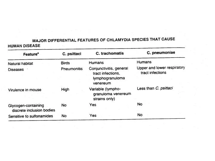
Epidemiology 1. Diseases - Adult inclusion conjunctivitis, infant pneumonia, non-gonococcal urethritis. 2. Transmission - Conjunctivitis, eye-to-eye by hand, droplets, flies, etc. Pneumonia, at birth via infected birth canal STD 3. Risk groups - Children in impoverished countries People with multiple sexual contacts 4. Geography - Conjunctivitis, endemic in Africa, Asia ansd South America Urogenital infections - world-wide
Mechanisms of Microbial Disease. 3rd Ed. 1998. M. Schaechter, et. al Eds.
Mechanisms of microbial Disease. 3rd ed. 1998. Schaechter, M. et. al. Eds