Download
1 / 1
10 likes | 96 Views
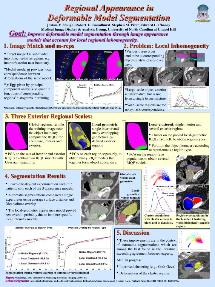
Regional Appearance in Deformable Model Segmentation. Joshua V. Stough, Robert. E. Broadhurst, Stephen M. Pizer, Edward L. Chaney Medical Image Display & Analysis Group, University of North Carolina at Chapel Hill.

E N D
Regional Appearance in Deformable Model Segmentation Joshua V. Stough, Robert. E. Broadhurst, Stephen M. Pizer, Edward L. Chaney Medical Image Display & Analysis Group, University of North Carolina at Chapel Hill Goal:Improve deformable model segmentation through image appearance models that account for local regional inhomogeneity. 1. Image Match and m-reps 2. Problem: Local Inhomogeneity • Various tissue types tend to be at corresponding object-relative places over days. Object 1 Object 2 • Target image I is subdivided into object-relative regions, e.g. interior/exterior near boundary. • Medial model m provides local correspondence between deformations of the same model. • p(I|m) given by principal component analysis on quantile functions of corresponding regions’ histograms in training. BladderProstate Bladder m-rep and surface with corresponding region highlighted RIQFs = Inverse CDFs Intensity histograms • Large-scale object-exterior is informative, but is not from a single tissue mixture. • Voxel-scale regions are too noisy, lack correspondence. • Regional intensity quantile functions (RIQFs) are amenable to Euclidean statistical methods like PCA. 3. Three Exterior Regional Scales: Global regions: sample the training image near the object boundary, compute the RIQFs for each case, interior and exterior. • Local-geometric: single interior and many overlapping geometrically-defined exterior regions • Local-clustered: single interior and several exterior regions • Cluster on the pooled local-geometric RIQFs (see left) to obtain region-types. • Partition the object boundary according to representative region-type. • PCA on the sets of interior and exterior RIQFs to obtain two RIQF models with Gaussian variability. • PCA on each region separately, to obtain many RIQF models that together form object appearance. • PCA on the region-type populations to obtain severalRIQF models. 4. Segmentation Results Global (red)versus local-geometric • Leave-one-day-out experiment on each of 5 patients with each of the 3 appearance models • Automatic segmentations compared a single expert rater using average surface distance and Dice volume overlap • The local-geometric appearance model proved best overall, probably due to its more specific local intensity models. Clusters in RIQF space. Local-geometric versus manual Cluster center densities. Cluster populations with cluster centers in black and as densities. Region-type partition for the bladder. Clustering yields biologically sensible regions. 5. Discussion • These improvements are in the context of automatic segmentations which are among the best found in the literature, exceeding agreement between experts. • Also, in progress: • Improved clustering (e.g., Gath-Geva) • Deformation of the cluster regions Segmentation trends, volume overlap of automatic versus manual Paper:Proceedings, 2007 Information Processing in Medical Imaging (IPMI ’07) Acknowledgments:Conceptual, algorithmic and code contributions from Joshua Levy, Gregg Tracton and Graham Gash. Partially funded by NIH-NIBIB P01 EB002779.