Download
1 / 3
30 likes | 119 Views
Dr. Deepak Talwar<br>Director & Chair, Pulmonary,<br>Sleep & Critical Care Medicine,<br>Metro Group of Hospitals, Noida<br>Dr. Deepak Talwar<br>Director & Chair, Pulmonary,<br>Sleep & Critical Care Medicine,<br>Metro Group of Hospitals, Noida

E N D
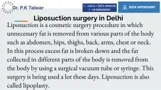
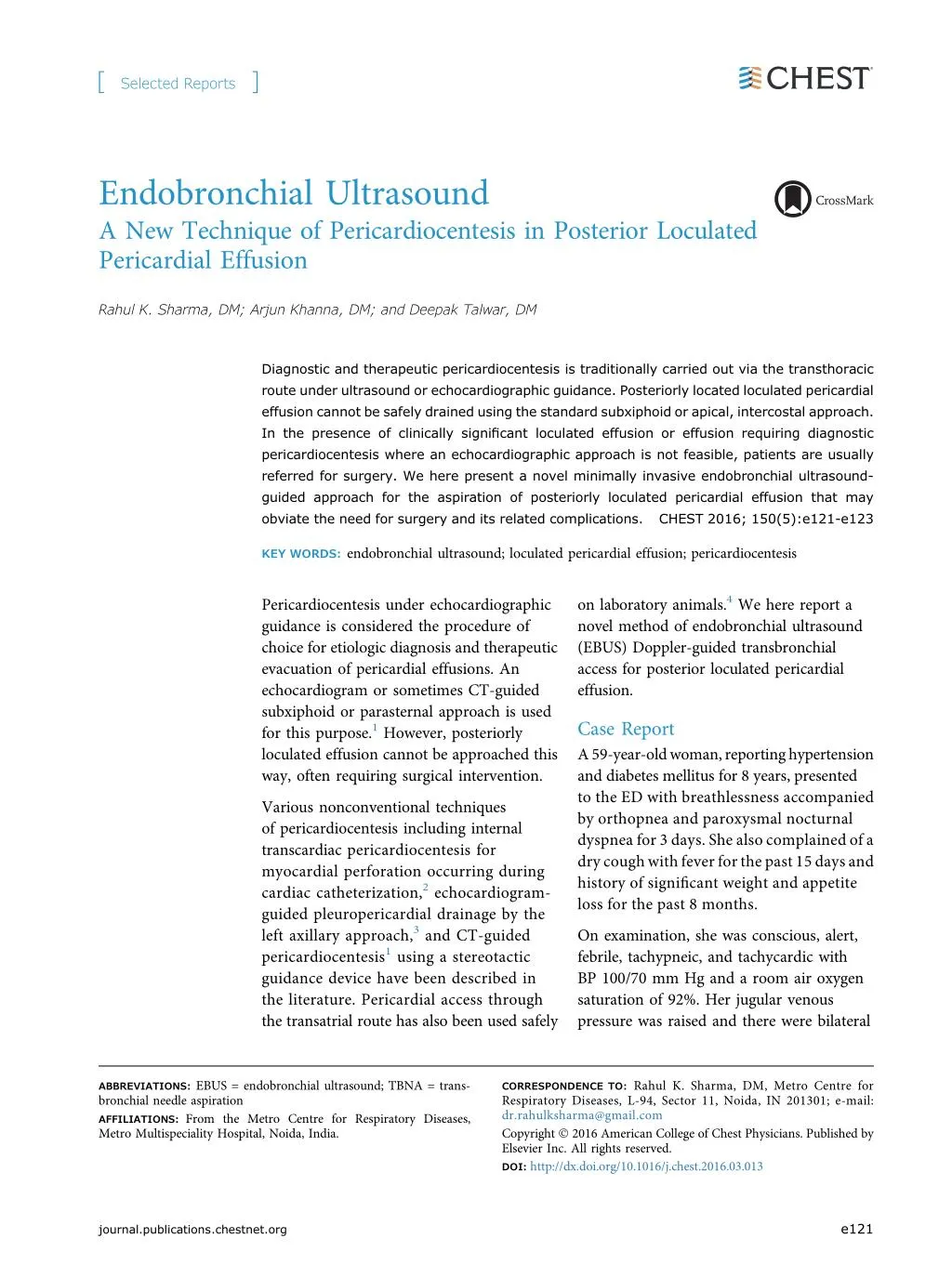
[ Selected Reports ] Endobronchial Ultrasound A New Technique of Pericardiocentesis in Posterior Loculated Pericardial Effusion Rahul K. Sharma, DM; Arjun Khanna, DM; and Deepak Talwar, DM Diagnostic and therapeutic pericardiocentesis is traditionally carried out via the transthoracic route under ultrasound or echocardiographic guidance. Posteriorly located loculated pericardial effusion cannot be safely drained using the standard subxiphoid or apical, intercostal approach. In the presence of clinically significant loculated effusion or effusion requiring diagnostic pericardiocentesis where an echocardiographic approach is not feasible, patients are usually referred for surgery. We here present a novel minimally invasive endobronchial ultrasound- guided approach for the aspiration of posteriorly loculated pericardial effusion that may obviate the need for surgery and its related complications. CHEST 2016; 150(5):e121-e123 KEY WORDS: endobronchial ultrasound; loculated pericardial effusion; pericardiocentesis on laboratory animals.4We here report a novel method of endobronchial ultrasound (EBUS) Doppler-guided transbronchial access for posterior loculated pericardial effusion. Pericardiocentesis under echocardiographic guidance is considered the procedure of choice for etiologic diagnosis and therapeutic evacuation of pericardial effusions. An echocardiogram or sometimes CT-guided subxiphoid or parasternal approach is used for this purpose.1However, posteriorly loculated effusion cannot be approached this way, often requiring surgical intervention. Case Report A59-year-oldwoman,reportinghypertension and diabetes mellitus for 8 years, presented to the ED with breathlessness accompanied by orthopnea and paroxysmal nocturnal dyspnea for 3 days. She also complained of a drycoughwithfeverforthepast15daysand history of significant weight and appetite loss for the past 8 months. Various nonconventional techniques of pericardiocentesis including internal transcardiac pericardiocentesis for myocardial perforation occurring during cardiac catheterization,2echocardiogram- guided pleuropericardial drainage by the left axillary approach,3and CT-guided pericardiocentesis1using a stereotactic guidance device have been described in the literature. Pericardial access through the transatrial route has also been used safely On examination, she was conscious, alert, febrile, tachypneic, and tachycardic with BP 100/70 mm Hg and a room air oxygen saturation of 92%. Her jugular venous pressure was raised and there were bilateral ABBREVIATIONS: EBUS = endobronchial ultrasound; TBNA = trans- bronchial needle aspiration AFFILIATIONS: From the Metro Centre for Respiratory Diseases, Metro Multispeciality Hospital, Noida, India. CORRESPONDENCE TO: Rahul K. Sharma, DM, Metro Centre for Respiratory Diseases, L-94, Sector 11, Noida, IN 201301; e-mail: dr.rahulksharma@gmail.com Copyright ? 2016 American College of Chest Physicians. Published by Elsevier Inc. All rights reserved. DOI: http://dx.doi.org/10.1016/j.chest.2016.03.013 e121 journal.publications.chestnet.org
Figure 1 – CT scan of the chest showing the (A) posteriorly loculated pericardial effusion and the (B) site of endobronchial ultrasound scope placement in the left lower bronchus for localization of pericardial effusion (arrow). crepitations and wheezing on chest examination, with no other systemic abnormality. and vital monitoring after informed consent. The subcarinal lymph node was approached from the right main bronchus and pericardial effusion was localized by an EBUS Doppler probe through the anterior wall of the left lower lobe bronchus. The pericardium was identified above the left atrium, which showed thickening and internal septations with no vascularity. Laboratory examination revealed hemoglobin 11.1 gm/dL, total leukocyte count 8,100/mm3, normal liver and kidney function tests, and raised brain natriuretic peptide levels (12,000 pg/mL). Workup for fever was negative. Chest radiograph showed cardiomegaly with increased vascular markings in parahilar region. Echocardiogram also revealed cardiomegaly with posteriorly located pericardial effusion, not amenable for echocardiogram-guided diagnostic aspiration. A CT scan of the chest confirmed the posteriorly located pericardial effusion with pretracheal and hilar mediastinal lymphadenopathy (Fig 1). Subsequently, an EBUS-TBNA needle was slowly advanced into the pericardial cavity (Fig 2). Ten milliliters of straw-colored pericardial fluid was aspirated and revealed lymphocytic exudative effusion with an adenosine deaminase level of 92 IU/L. The subcarinal lymph node showed necrotizing granuloma, confirming the diagnosis of TB. The patient tolerated the procedure well with no postprocedural complications. She was subsequently started on anti-TB therapy with steroids in view of pericardial involvement being diagnosed by EBUS-guided pericardiocentesis; A diagnosis of pulmonary TB was suspected clinically, and an EBUS-transbronchial needle aspiration (TBNA) was done under conscious sedation with electrocardiographic Figure 2 – Endobronchial ultrasound images showing a transbronchial needle aspiration needle in the pericardial space, with the left atrium (LA) below showing widening of pericardial space during atrial contraction. [ 1 5 0 # 5 C H E S T N O V E M B E R 2 0 1 6 ] e122 Selected Reports
she has improved symptomatically on follow-up at 4 weeks. experience of EBUS-TBNA of mediastinal lymph nodes, studies have shown that it is a safe procedure with very low complication rate.7 Discussion Diagnostic pericardiocentesis is traditionally carried out via echocardiogram-guided transthoracic route, but most posteriorly located pericardial effusions cannot be drained using this technique, necessitating the use of surgery in such cases. A previous study describes a bronchoscopic approach to pericardial effusion in three patients by gaining needle access through the left lower lobe bronchus or through the distal trachea based on CT assessment.5This was somewhat of a blind procedure, which though uneventful in the present case series, has potential to cause nearby vascular injury. This technique is rather easy for operators skilled in TBNA, and is safe, economical, and well-tolerated. To the best of our knowledge, this is the first such report of its kind from the Indian subcontinent. Though not adequately researched, it forms a landmark in the management of posterior pericardial effusion and requires further study to understand the benefits and possible hazards of this procedure. Acknowledgments Financial/nonfinancial disclosure: None declared. References 1. Duvernoy O, Magnusson A. CT-guided pericardiocentesis. Acta Radiol. 1996;37(5):775-778. 2. Fisher JD, Kim SG, Ferrik KJ, et al. Internal transcardiac pericardiocentesis for acute tamponade. Am J Cardiol. 2000; 86(12):1388-1389. 3. De Divitiis M, Dialetto G, Covino FE, et al. An unusual procedure for the treatment of simultaneous pericardial and pleural effusions. G Ital Cardiol. 1999;29(7):796-798. 4. Verrier RL, Waxman S, Lovett EG, et al. Transatrial access to the normal pericardial space: a novel approach for diagnostic sampling, pericardiocentesis, and therapeutic interventions. Circulation. 1998;98(21):2331-2333. 5. Ceron L, Manzato M, Mazzaro F, et al. A new diagnostic and therapeutic approach to pericardial effusion: transbronchial needle aspiration. Chest. 2003;123(5):1753-1758. 6. Gella V, Ghana S, Srinivas U. Concurrent diagnostic pericardiocentesis and subcarinal mediastinal lymph node [abstract]. Eur Respir J. 2015;46(suppl 59):PA782. 7. Asano FL, Aoe M, Ohsaki Y, et al. Complications associated with endobronchial ultrasound-guided transbronchial needle aspiration: a nationwide survey by the Japan Society for Respiratory Endoscopy. Respir Res. 2013;14:50. We describe a novel real-time, EBUS-guided approach to drain the posteriorly located pericardial effusion. This is a simple and safe way of sampling even minimal effusions for diagnostic purposes. The pericardial cavity can be safely reached with accuracy under real-time Doppler imaging without any risk of touching the myocardium. This approach can be used to establish an otherwise difficult diagnosis of pericardial diseases, including the staging of tumors in patients with a malignancy and pericardial effusion.6It could also represent a less invasive alternative to surgery when evacuation cannot be performed via either subxiphoid or parasternal puncture. In theory, it is possible that gaining access to the pericardium through the nonsterile bronchus could lead to pericardial infection. No published literature is available on this issue; nevertheless, looking into the e123 journal.publications.chestnet.org