Download
1 / 47
470 likes | 499 Views
DR.DUAA HIASAT. Osteoporosis. To define osteoporosis. To know the causes & risk factors of osteoporosis. To know when to screen for osteoporosis. To talk about preventive measures of osteoporosis. To recognise general principles of management. Objectives :.

E N D
DR.DUAA HIASAT Osteoporosis
To define osteoporosis. To know the causes & risk factors of osteoporosis. To know when to screen for osteoporosis. To talk about preventive measures of osteoporosis. To recognise general principles of management. Objectives :
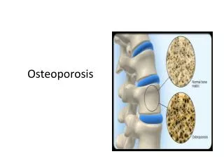
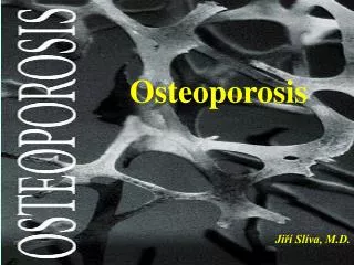
A skeletal disorder characterised by decreasedbone strength ( density and/or quality) leading to increased risk for fragility fracture. Bones of hip,spine and wrist most commonly affected. What is osteoporosis??
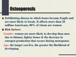
Osteoporosis is a major public health problem, which results in substationalmorbidity,mortality and high costs. Osteoporosis is an extremely serious disease and is not part of the natural aging process. Why is it important ??
Maximum bone density is attained in young adulthood. Women then typically lose 50% bone mass over their remaining lifetime. Bone loss begins in the premenopausal years & accelerates in the early postmenopausal years. Lack of estrogen causes a release of osteoclastic inhibition leads to imbalance b/w bone resorption and formation. Pathogenesis :
1 – Idiopathic age related osteoporosis (most common). 2- Secondary osteoporosis. Causes of osteoporosis:
Osteoporosis secondary to disease states: 1. Metabolic conditions: Ex: Calcium deficiency ,Vit.D deficiency, Malnutrition,Idiopathichypercalciurea,Scurvy. 2. Endocrine conditions : Ex:Thyrotoxicosis,Cushings,Hypogonadism Prolactinoma,HyperPTH,Hypoamenorrheic female runners (Athletes triad). Cont…
3. Gastrointestinal disease: Ex:IBD,Malabsorption (e.g Celiac sprue). 4. Bone marrow infiltration : Ex: Lymphoma,Leukemia,Multiple myeloma. 5. Drugs : Ex: Anticonvulsants,Thyroid hormones ,PPIs,SSRIs, Corticosteroids ( 7.5 mg/d/3 mo),GnRH agonists, Chronic heparin/warfarintherapy,furosemide,Lithium. 6. Lifestyle: Ex: Nutrition ,Alcohol,Smoking ,Immobilazion, Inactivity. 7. Others : Rhematoidarthritis,COPD.
Trabecular bone more commonly affected than compact bone. Spine ( vertebral column),pelvis,hip ( femoral neck),distal radius. Organs involved
Majors risk factors for osteoporosis : Age ≥ 65 years. Gender ( more in females). White or Asian race. Menopausal status. Personal history of fracture as an adult (esp. older than 45 years ; not including fingers,toes, and skull). History of fragility fracture in a first-degree relative (esp.maternal hip fracture). Current smoking. Low body weight ( BMI <21 kg/m2). Use of oral steroids > 3 months. Who is at risk??
Additional risk factors: Estrogen deficiency at an early age (<45 years). Low physical activity. Excessive alcohol (more than 2 drinks/day Poor health.
It’s usually asymptomatic until fracture occurs. Loss of height is common. Spontaneous fracture. Collapse of vertebrae. Clinical manifestations of osteoporosis:
1- Based on bone densometry ( DEXA scan) with the use of T or Z score. 2- Clinical diagnosis if fragility fracture regardless of T/Z score. Diagnosis :
Central DEXA is the gold standard for assessment of BMD. -Two beams of different energy are directed at The patient and the difference in absorption rate by the patient body is recorded to quantify the amount of BMD. Cont…
T–score is bone density expressed as number of standard deviations above or below mean BMD value for a normal young adult measured by DEXA. WHO criteria for diagnosis of bone status are : T-score -1 is normal. T-score between -1 and -2.5 is osteoponia. T-score ≤ -2.5 is osteoporosis. T-score ≤ -2.5 + fractue is sever osteoporosis. T and Z score
Z-score is bone density expressed as number of SDs from normal mean value for age,sex and ethnicity/race. Z-score > -2 is normal. Z-score ≤ -2 is osteoporosis. In premenopausal women,men <50 years old, and children,use Z-score instead of T-score. Cont..
National Osteoporosis Foundation (NOF) recommends that all women 65 years should be screened regardlessof the presence or absence of risk factors. Younger postmenopausal women with one or more risk factors (other than being white,postmenopausal,and female) should also be screened for osteoporosis. When to do bone densometry??
U.S Preventive Services Task Force (USPSTF) also recommends universal BMD screening for all women ≥ 65 years and for women ≥ 60 years ifthere are risk factors for osteoporotic fractures. In contrast to NOF, the USPSTF makes no recommendation for or against routine screening in postmenopausal women younger than age 60 years or aged 60 to 64 years without increased risk for osteoporotic fracture.
It’s the least costly approach to osteoporosis and should begin in youth . Prevention:
All patients being considered for pharmacologic therapy should be counseled on the importance of calcium,vitaminD,weight-bearing exercise and fall preventive strategies to further reduce their risk of fracture. Before initiating pharmacologic treatment patients should also be evaluated for secondary causes of osteoporosis. Treatment :
NOF recommends treatment for : Patients with T-score below -2. Patients with T-score below -1.5 if one or more risk factors are present. Patients who already have had osteoporotic fracture (s). Who should be treated?
FDA – Approved therapeutic options A) Prevention (stops bone loss) : Estrogen. Bisphosphonates ( Alendronate,Risedronate, Ibandronate). SERM (Raloxifne). B) Treatment ( reduces spine fractures): Calcitonin. Parathyroid hormone. Cont..
Bishosphanates: The most commonly used therapies for osteoporosis.They are potent inhibitors of bone resorption. Alendronate was the first FDA-Approved drug , it’s taken orally 5 mg daily or 35 mg weekly for prevention ,10 mg daily or 70 mg weekly for treatment. It reduces the incidence of vertbral and non-vertebral fractures by about half.
Because a minority of patients may suffer from erosive esophagitis ,the patient should be advised to take it on empty stomach with water upon awakening ,remain upright ,and avoid food for 30 minutes afterwards. Other Side effects: • GERD ( common). • Myalgia,arthralgia. • Jaw neck necrosis (rare but serious ). • Atrial fibrillation. Cont..
Estrogen therapy alone or with a progestin is indicated for the prevention of osteoporosis only. However, because of the potential risks of estrogen, the FDA recommends that if a woman doesn’t have menopausal symptoms, nonestrogentreaments should be carefully considered before using estrogen therapy solely for the prevention of postmenopausal osteoporosis. Estrogen:
It selectively binds to estrogen receptors and inhibits bone resorption. Raloxifene is FDA-Approved for both the prevention and the treatment of osteoporosis. Although Raloxifene decreases the risk of vertebral fractures,there is no evidence that it decreases the risk of nonvertebral fractures including the hip. SERM:
It remains unclear whether anti-fracture efficacy is sustained beyond 3-5 yrs. Stopping treatment is followed by increased remodeling,bone loss and further structural damage. Bone loss is likely to recur sooner after cessation of HRT or raloxifene than bisphosphonates. How long should treatment continue?
It is generally recommended tht central DEXA be repeated no less than 2 years from initiation or change in drug therapy to detect significant changes in BMD. It is also important to note that drug therapy may decrease fracture risk without an apparent increase in BMD. When we should re-evaluate ??
If BMD is > -1 SD do not treat. If BMD is -1 to -2.5 SD treat if fracture is present. If BMD is <-2.5 SD treat whether or not a fracture is present. In conclusion
A 61-year old white postmenopausal woman comes to your office for routine health examination.She has a history of osteoarthritis , she smokes 1 pack of cigarettes Per day.She fractured her left wrist at age of 50 after falling down some stairs.Her diet is low at calcium-rich food.She is on no medication except calcium.Her physical examination is normal. Case 1
You believe that she is at risk for osteoporosis the test of choice is ? A.Ultrasound B.Peripheral DEXA. C.Central DEXA. D.Plain X-ray. E.Quantitative CT scan. Q.1
You order a central DEXA scan to the patient and scan returns with T score of -1.3 for the lumbar spine and -1.9 for the total hip.What do you recommend to the patient at this time? A.No action needed. B.Repeat DEXA in 6 months. C.Recommend adequate Ca intake, weight bearing exercise . D.Recommend adequate Ca intake, weight bearing exercise and bisphosphonates. E.Recommend adequate Ca intake , weight bearing exercise and calcitonin. Q.2
Which of the following is not an established major risk factor for osteoporosis? A.Low body weight B.Current smoking. C.History of fragility fracture in 1st degree relative. D.Low calcium intake. E.Chronic use of steroids. q.3
Which of the following is in not an associated risk factor for osteoporosis? A.COPD B.HyperPTH. C.Cigarette smoking. D.Obesity. E.Excessive alcohol intake. q.4
What is the most common presenting fracture in osteoporosis? A.Wrist fracture (Colles fracture). B.Vertebral compression fracure. C.Femoral neck fracture. D.Tibial fracture. E.Femoral head fracture. q.5
Which of the following sites for osteoporotic fracure is most commonly associated with morbidity and mortality? A.Ward’s triangle (hip). B.Femoral neck ( hip). C.Thoracic vertebrae (spine). D.Lumbar vertebrae ( spine). E.Distal radius (wrist). q.6
Which of the following is not recommended for treatment of established osteoporosis? A.Estrogen. B.Calcium and Vitamin D. C.Bisphosphonates. D.SERMs. E.Calcitonin. q.8
Which of the following studies may be indicated in an asymptomatic patient recently diagnosed with osteoporosis? A.24-hr urine calcium. B.Serum 25-hydroxy vitamin D. c.PTH. D.TSH. E.All of the above. q.9
A 32 years old woman is seeing because her mother as been diagnosed with osteoporosis.She asks you what type of exercise will help her prevent the development of the disease.According to the recommendations,which of the following excercises is most appropriate to help her maintain bone mass?? A.Tennis. B.Swimming. C.Cycling. D.Skating. E.Skiing. Case 2
You are treating an elderly postmenopausal woman with osteoporosis . She recently suffered an acute osteoporotic vertebral fracture and is suffering from secondary pain.Which of the following treamentsalo has an analgesic effects with respect to bone pain? A.Estrogen. B.Combination of calcium & Vitamin D. C.Calcitonin. D.Alendronate. E.Raloxifene. Case 3
You have just diagnosed osteoporosis in a postmenopausal woman.She is considering treatment alternatives and wonders about the bisphosphonates.Which of the following is the best description of how this class of medication works? A.They increase calcium absorption in the GI tract. B.They block the activity of the cytokines that stimulates bone reabsorption. C.They bind to bone surfaces to inhibit osteoclast activity. D.They stimulate osteoblasts and increase bone formation. E.They mimic estrogen’s effect on bone. Case 4
Case 1 Q1 c Q4 d Q7 a Q2 d Q5 b Q8 a Q3 d Q6 b Q9 e CASE 2 answer is a CASE 3 answer is c CASE 4 answer is c Answers: