Download
1 / 36
360 likes | 1.08k Views
Management of large rectal adenoma. Dr. Hester YS Cheung. Department of surgery Pamela Youde Nethersole Eastern Hospital. Adenoma. Neoplastic polyps Precursor of colorectal cancer. Anatomical distribution. National polyp study Colonoscopy study 8% in rectum. O’Brien et al. 1990.

E N D
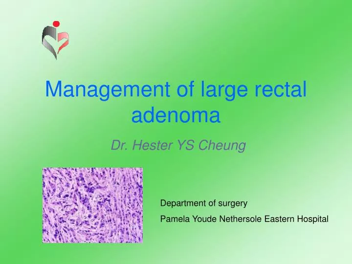
Management of large rectal adenoma Dr. Hester YS Cheung Department of surgery Pamela Youde Nethersole Eastern Hospital
Adenoma • Neoplastic polyps • Precursor of colorectal cancer
Anatomical distribution • National polyp study • Colonoscopy study • 8% in rectum O’Brien et al. 1990
Large rectal adenoma • Large polyps More than 30mm in size The larger the size, the greater is the risk of malignancy Williams 1999
Malignancy risk Shinya and Wolff 1979
Management • Diagnosis • Work-up • Surgical treatment • Follow-up
Diagnosis • Rectal examination • Rigid sigmoidoscopy • Flexible sigmoidoscopy
Diagnosis • Rigid sigmoidoscopy • Villous adenoma • 97% within 30cm from anal verge • Problems • View obscured by blood or mucus • Sub-optimal insufflation
Diagnosis • Flexible sigmoidoscopy • Advantages • Possible to negotiate the rectosigmoid junction and pass up to splenic flexure • Relatively comfortable • Polypectomy
Diagnosis • Flexible sigmoidoscopy Yield is three times as high as with the rigid instrument Marks et al. 1979 McCallum et al. 1984
Work-up • Biopsy • Colonoscopy • Synchronous polyps(20-26%) / cancer(3%) • Endoluminal ultrasound • Mid and low rectal polyps : below 12cm
Work-up • Endoluminal ultrasound • Depth of rectal wall invasion • T- and N-stages, if malignant • Guides further management
Surgical treatment • Endoscopic polypectomy • Peranal excision • Perineal surgical manoeuvre • Abdominal procedures • Others
Endoscopic polypectomy • Ideal for • Small polyps • Larger polyp with a stalk • Sessile polyps • Piecemeal removal
Endoscopic mucosal resection (EMR) • Colonoscopy using electrocautery • Reported by Deyhle et al. 1973 • Early gastric cancer by Tada et al. • Indications • Flat-type or depressed lesions
Positioning Injection EMR Wiring Excision Extraction Koji Matsuda Gastrointestinal endoscopy 2001
Peranal excision • Large polyp with a long pedicle in lower rectum • Digitally palpable • Polyp hooked down through the anal orifice • Pedicle transfixed and excised • 10-15mm margin
Perineal surgical manoeuvre • Not amenable to endoscopic polypectomy • Too large and sessile • Behind a fold • Too low
Perineal surgical manoeuvre • Conventional transanal excision (Park’s approach) • Transanal endoscopic microsurgery (TEM) • Trans-sphincteric excision
Conventional approach (Park’s) • Low rectal adenoma (digitally palpable) • Lithotomy / Jack-knife position • Submucosal plane infiltration with saline and adrenaline • 1cm margin • Submucosal excision
Transanal endoscopic microsurgery (TEM) • First clinical application in 1983 • Complex • Costly • Needs substantial training • Conglomeration of endoscopic and laparoscopic technique Buess et al. 1984 Buess, 1994
TEM • Indications • Upper and middle rectal lesions • Primarily for benign adenoma • Local excision for cancer palliation
TEM • Depth of excision
TEM • Benefits • For removal of villous adenomas that cannot be removed by conventional technique • Up to 24cm from anal verge Buess 1992
Complications Hemorrhage Perforation Incontinence Rectal stricture Suture dehiscence Urinary tract infection Urinary retention TEM
Results • Transanal endoscopic microsurgery
Trans-sphincteric excision • Originally described by Bevan • Revived by York Mason • Indications • For anterior or anterolateral lesions 8-12cm from the anal verge • Poor risk patients who cannot withstand major laparotomy Bevan 1917 Mason 1970
Trans-sphincteric excision • Anal sphincters and rectal wall divided in the longitudinal axis • Sphincter function retained if the cut layers are sutured accurately
Trans-sphincteric excision • Advantages Too high for transanal excision Under direct vision Lower risks of perforation Tumor upper limit can be reached more easily
Trans-sphincteric excision • Disadvantages Inferior function results Higher morbidity Replaced by TEM or laparoscopic approach
Abdominal procedures • Radical surgery • Indications • Upper and mid-rectal lesions (TEM not available) • Lesions behind a mucosal fold • Approach • Anterior / low anterior resection • Laparoscopic approach
Other techniques • Diathermy fulguration • Endoscopic transanal resection of tumor • Laser photocoagulation • Photodynamic therapy
Other techniques • Disadvantages • No intact specimen for accurate histological examination and staging For palliation in poor risk patients