Download
1 / 66
700 likes | 815 Views
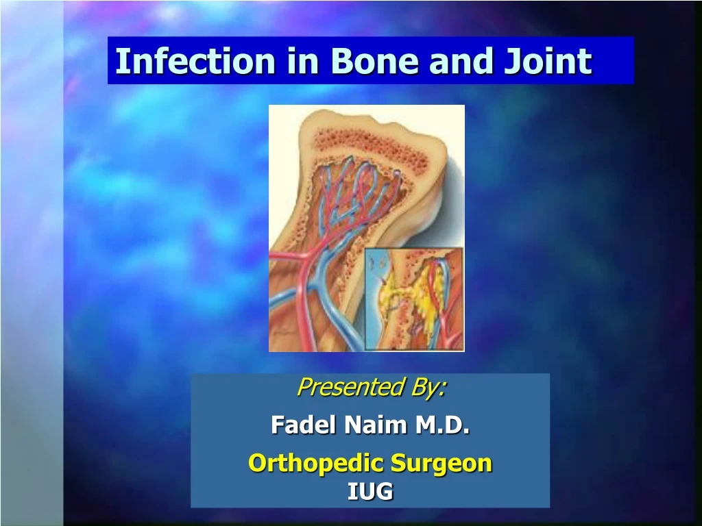
Infection in Bone and Joint. Presented By: Fadel Naim M.D. Orthopedic Surgeon IUG. Osteomyelitis. osteon (bone) myelo (marrow) itis (inflammation) Inflammation and destruction of bone caused by aerobic and anaerobic bacteria, mycobacteria , and fungi.

E N D
Infection in Bone and Joint Presented By: FadelNaim M.D. Orthopedic Surgeon IUG
Osteomyelitis • osteon (bone) • myelo (marrow) • itis (inflammation) • Inflammation and destruction of bone caused by aerobic and anaerobic bacteria, mycobacteria, and fungi • The term osteomyelitis does not specify the causative organism or the disease process
Epidemiology • Common in young children • Common with malnutrition, immunodeficiency - with decreased resistance of the patient • Boys> girls • History of trauma • Decreasing in incidence & severity & mortality with advent of newer antibiotics
Osteomyelitis Classification: • Duration Acute, Subacute or Chronic • Route of infection Hematogenous or Exogenous • Host response Pyogenic or Granulomatous
Source Of Infection • Hematogenous spread • Direct inoculation • Contiguous focus of infection
The most common site is the rapidly growing and highly vascular metaphysis of growing bones • The apparent slowing or sludging of blood flow as the vessels make sharp angles at the distal metaphysis • predisposes the vessels to thrombosis • the bone itself to localized necrosis and bacterial seeding
The joint is usually spared from infection unless the metaphysis is intracapsular, as is found in the proximal part of: • The radius • The humerus • The femur
Age variation Neonates: • Extensive bone necrosis • Increased ability to absorb large sequestrum • Increased ability to remodel • Epiphysio-metaphyseal vascular connection • leading to secondary septic arthritis
Age variation Adults: • No subperiosteal abscess due to adherent periosteum • Soft tissue abscess • Vascular connection with the joint leading to secondary septic arthritis
Clinical Pictures • Pain, restless • Malaise and fever • The limb is held still (pseudo paralysis) • Sometimes mild or absent (neonates)
Acute OsteomyelitisDiagnosis • History and clinical examination • CBC, ESR, B.C. • X-ray (normal in the first (10-14) days • Ultrasound • Bone Scan Tc 99, Gallium 67 • MRI • Aspiration
Radiographic Findings • Usually reflect the destructive process but lag at least two weeks behind the process of infection • The earliest changes are: • Swelling of the soft tissue • Periosteal thickening and/or elevation • Focal osteopenia • At least 50% to 75% of the bone matrix must be destroyed before radiographs show lytic changes
(A) Proximal humerus at day 1 of infection - no visible changes. • (B) Proximal humerus at day 12 of infection
Plain-film radiograph showing osteomyelitis of the second metacarpal (arrow). Periosteal elevation, cortical disruption and medullary involvement are present.
The above X-ray of the left ankle of a 10-year-old boy shows lucency in the tibialmetaphysis secondary to acute hematogenousosteomyelitis (AHO). The above X-ray of the right ankle of a 10-year-old boy shows lucency in the tibialmetaphysis secondary to acute hematogenousosteomyelitis (AHO).
Here is an X-ray of an AHO lesion extending into the growth plate
Radiological studies • MRI : Early detection and surgical localization of osteomyelitis. Sensitivity ranges from 90-100%. • Radionuclide bone scanning : A 3-phase bone scan with technetium 99m is probably the initial imaging modality of choice Show increase activity but it is a non specific sign of inflamation.
This MRI sagittal section shows the same AHO lesions with the right lesion extending into the growth plate.
Bone scans, both anterior (A) and lateral (B), showing the accumulation of radioactive tracer at the right ankle (arrow). This focal accumulation is characteristic of osteomyelitis.
Labratory • Theleukocyte count (WBC), erythrocyte sedimentation rate (ESR), and C-reactiveprotein level (CRP) should be monitored • At the time of admission • During treatment • During follow-up • In all patients with osteomyelitis on a weekly basis
Diagnosis requires 2 of the 4 following criteria: • Purulent material on aspiration of affected bone • Positive findings of bone tissue or blood culture • Localized classic physical findings of bony tenderness, with overlying soft-tissue erythema or edema • Positive radiological imaging study
Differential Diagnosis • Acute Septic Arthritis • Acute monoarticular rheumatoid arthritis • Sickle cell crisis • Cellulitis • Ewing’s Sarcoma
Complications • Septicemia & metastatic abscesses • Septic arthritis • Growth disturbance (children) • Pathological fracture • Chronic osteomyelitis
SubacuteOsteomyelitis • Results from a less virulent Microorganism, or a patient with an elevated resistance. • Occurs Mostly at the Distal Femur or Proximal Tibia • On X-Ray: Brodie’sAbcess • Small and Oval in shape • It is surrounded by sclerotic bone • May be mistaken for OstieoidOsteoma
SubacuteOsteomyelitis • An image depicting subacute osteomyelitis
Chronic Osteomyelitis • The coexistence of infected, nonviable tissues and an ineffective host response leads to the chronicity of the disease
Chronic Osteomyelitis Factors responsible for chronicity • Local factors:Cavity, Sequestrum, Sinus, Foreign body, Degree of bone necrosis • General: Nutritional status of the involved tissues, vascular disease, DM, low immunity • Organism: Virulence • Treatment: Appropriateness and compliance • Risk factors: Penetrating trauma, prosthesis, Animal bite
Pathologic features of chronic osteomyelitis • Sequestrum: When both the medullary and the periosteal blood supplies are compromised, large areas of dead bone (sequestra) may be formed • Involucrum: New bone forms from the surviving fragments of periosteum and endosteumin the region of the infection to form an encasing sheath of live bone • sinus tract: A bone cavity may persist or the space may be filled with fibrous tissue, which may connect with the skin surface by the sinus tract
Chronic Osteomyelitis Types • A complication of acute Osteomyelitis • Post traumatic • Post operative
Chronic Osteomyelitis • Low gradefever, if present • ESR usually elevated, reflecting chronic inflammation • Theblood leukocyte count ( WBC ) is usually normal • If a sinustract becomes obstructed, the patient may present with a localizedabscess and/or an acute soft-tissue infection
Chronic Osteomyelitis Organism • Usually mixed infection • Mostly staph. Aureus E. Coli . strep pyogen, proteus
Treatment of Osteomyelitis • A close interaction between various specialists is important to improve the management of this disease • Orthopaedic surgeons • Plastic and vascular surgeons • Infectious disease specialists
Treatment of Osteomyelitis • Adequate drainage • Thorough débridement • Obliteration of dead space • Wound protection • Specific antimicrobial coverage • Correcttion of host defects • Improving the nutritional, medical, and vascular status of the patient • Good nutrition • Smoking cessation • Control of specific diseases such as diabetes
Bone Stabilization • If skeletal instability is presentmeasures must be taken to achieve stability with plates, screws, rods, and/or an external fixator. • External fixation is preferred over internal fixation. • Ilizarov external fixation: • allows reconstruction of segmental defects and difficult infected nonunion. • An extended period of treatment with the device, averaging 8.5 months. • The sites of the wires or pins usually become infected and the device is painful.
Soft-tissue Coverage • Adequate soft-tissue coverage of the bone is necessary to arrest osteomyelitis • Small soft-tissue defects may be covered with a split-thickness skin graft • For large soft-tissue defects or an inadequate soft-tissue envelope, local muscle flaps and free vascularized muscle flaps may be placed in one or two stages • Healing by so-called secondary intention should be discouraged
Septic Arthritis • Septic arthritis : • Direct invasion of joint space by a variety of microorganisms, including a variety of bacteria, viruses, mycobacteria, and fungi. • Reactive arthritis: • A sterile inflammatory process, may be the consequence of an infectious process located somewhere else in the body.
Septic Arthritis • 50% of cases in children <3 years • The hip joint is the common site in <3years, whereas the knee joint is more common in older children.
7.8 cases per 100,000 person-years • The incidence of gonococcal arthritis is 2.8 cases per 100,000 person-years • Septic arthritis is becoming increasingly common among people who are immunosuppressed and elderly people who have a variety of co-morbid diagnoses
Most of these infections occur in very young and very old people and among people who abuse intravenous drugs • The most commonly involved joint: • Knee (50%) • Hip (20%) • Shoulder (8%) • Ankle, and wrists (7% each) • Elbow, interphalangeal, sternoclavicular, and sacroiliac joints each make up 1-4%of cases