Download
1 / 57
630 likes | 1.15k Views
Edema. Increased water in brain parenchyma. . Types of Herniations. Subfalcine herniation (cingulate gyrus) Transtentorial Herniation (uncal gyral) Tonsillar Herniation. Mass Tumor Blood clot Abscess Local edema. S. T. Transtentorial Herniation [involve Uncal gyral].

E N D
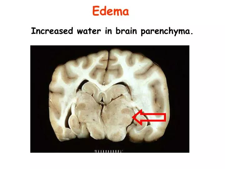
Edema Increased water in brain parenchyma.
Types of Herniations • Subfalcine herniation (cingulate gyrus) • Transtentorial Herniation (uncal gyral) • Tonsillar Herniation
Mass • Tumor • Blood clot • Abscess • Local edema S T
Transtentorial Herniation [involve Uncal gyral]
Transtentorial Herniation [Uncus]
Cerebral HerniationComplication of Intracranial Hypertension • Tonsillar herniation • Cerebellar tonsils herniate into the foramen magnum. • Causes "coning" of the cerebellar tonsils • Produces cardiorespiratory arrest • Coma and Death
Complications of Tonsillar Herniation & Increased ICP • Hemorrhagic lesion of the mid Brain and Pons : Secondary Brain stem or Duret hemorrhage • Linear hemorrhage
Duret hemorrhage: pathogenesis • Kinking of the penetrating median and paramedian pontine arteries that branch off the basilar artery.
Duret hemorrhage : causes • Tonsillar Herniation • Intracranial Neoplasm • Intracranial hemorrhage (basal ganglia )
HydrocephalusAccumulation of excessive CSF within the ventricular system & enlargement
Communicating Hydrocephalous. • Meningitis. • Subarachnoid hemorrhage Obstruction in subarachnoid space
Non communicating Hydrocephalous (Obstructive) • Medulloblastoma, • Ependymoma No communication between ventricles and subarachonoid space.
Congenital hydrocephalous (present at birth) • Aqueductal stenosis (narrowing) is the most frequent cause. • Blockage of fourth ventricle outlet (Dandy Walker Syndrome) – due to congenital malformation
The gross shows hydrocephalus of the frontal horns of the lateral ventricles.
Hydrocephalous before the fusion of the cranial Sutures [Head circumference increase]
Hydrocephalous after the fusion of the Sutures, produce Ventricular expansion and Increased Intracranial Pressure
Anencephaly 20
Types of spina bifida: A: Spina bifida occultaB: MeningoceleC: Meningomyelocele Spina bifida 21
Syringomyelia Note the collapsed cystic cavity (syrinx) in the center of the cervical spinal cord 22
Brain contusion The contrecoup injury involves the frontal and temporal lobes (left arrows) The coup lesion (site of impact) involves the cerebellum (right arrow). 23
Red Neuron 25
Border zone infarct: Watershed infarct : Follows a Hypotensive episode. Lesion lies at the boundary between the anterior and middle cerebral artery territories.
Lacunar Infarcts The arrows show multiple small cystic spaces (liquefactive necrosis) that are most prominent in the basal ganglia. 33
Lacunar infarcts Cause : Chronic hypertension Site: The pons.
Microscopically, a neutrophilic exudate is seen involving the meninges
Spongiform encephalopathy of gray matter : brain lesion in CJD
Complications: sequel Edema can lead to herniation and death. Communicating hydrocephalous.
Meningeal Syphilis 1 of 2 Neurosyphilis is a tertiary stage of syphilis – only in 10% with untreated syphilis May involve spinal Meninges: produce thickening. Produce meningeal fibrosis and secondary Hydrocephalous.
Atrophy There is marked atrophy seen superiorly and laterally.
The cortical atrophy leads to compensatory dilation of the cerebral ventricles [hydrocephalus ex vacuo ]
Self Study For anatomy, not for exam