Download
1 / 36
360 likes | 511 Views
Carbohydrates. Part 2. Signs and symptoms of DM. Polydipsia (excessive thirst), Polyphagia (increased food intake), Polyuria (excessive urine production), Rapid weight loss, Hyperventilation, Mental confusion,. Complications of DM. Microvascular problems such as: nephropathy,

E N D
Carbohydrates Part 2
Signs and symptoms of DM • Polydipsia (excessive thirst), • Polyphagia (increased food intake), • Polyuria (excessive urine production), • Rapid weight loss, • Hyperventilation, • Mental confusion, M. Zaharna Clin. Chem. 2009
Complications of DM • Microvascular problems such as: • nephropathy, • neuropathy, • and retinopathy. • Macrovascular problems • Increased heart disease is also found in patients with diabetes. M. Zaharna Clin. Chem. 2009
Pathophysiology of Diabetes Mellitus • Type 1 and Type 2 diabetes: there is an increase in blood glucose levels (hyperglycemic). • There is also elevation of glucose in urine (glucosuria) if glucose levels in blood exceeds 180 mg/dl. M. Zaharna Clin. Chem. 2009
Ketoacidosis • The individual with type 1 diabetes has a higher tendency to produce ketones. • Absence of insulin and with increased glucagon leads to gluconeogenesis and lipolysis. • The liver thus produces large amounts of ketone bodies, which are moderately strong acids. • The result is severe acidosis • the decrease in pH impairs tissue function, most importantly in the central nervous system. M. Zaharna Clin. Chem. 2009
Hyperosmolar Nonketonic States • Type 2: have very little ketone production, but have a greater tendency to develop hyperosmolar nonketonic states. • This disorder is caused by elevated blood sugar levels and is usually brought on by a coexisting condition, such as an illness or infection. • can be a life-threatening emergency M. Zaharna Clin. Chem. 2009
Hyperosmolar Nonketonic States • Criteria for hyperosmolar nonketonic states include: • serum osmolality of 320 mOsm/kg (275-299) • plasma glucose level greater than 600 mg/dL, • intense dehydration, • no ketoacidosis, • Hyperglycemia and the rise in concentration of plasma proteins that follow intravascular water loss cause a hyperosmolar state. M. Zaharna Clin. Chem. 2009
Hyperosmolar Nonketonic States • In the presence of a hyperglycemic, hyperosmolar state, if the renal water loss is not compensated by oral water intake, then hypovolemia follows dehydration. • Hypovolemia, in turn, leads to hypotension, and hypotension results in impaired tissue perfusion. • Coma is the end stage of this hyperglycemic process, when severe electrolyte disturbances occur in association with hypotension. • Ketones are not observed because glucagon is not able to stimulate lipolysis. M. Zaharna Clin. Chem. 2009
Criteria for Testing for Prediabetes and Diabetes • Forms of impaired glucose metabolism that do not meet the criteria for diabetes mellitus include impaired fasting glucose and impaired glucose tolerance. • These have a relatively high risk for the development of diabetes • First, those patients with: • fasting glucose levels ≥100 mg/dL but <126 mg/dL were called the impaired fasting glucose group. • Another set of patients who had 2-hour OGTT levels of ≥ 140 mg/dL but <200 mg/dL was defined as having impaired glucose tolerance. M. Zaharna Clin. Chem. 2009
Risk Factors for Diabetes • According to ADA recommendations, all adults older than 45 years should have a measurement of fasting blood glucose every 3 years unless the individual is otherwise diagnosed with diabetes. • Testing should be carried out at an earlier age or more frequently in individuals additional risk factors, as follows: • Obesity • Family history in 1st degree relative • History of GDM or > 4.1 Kg baby • Hypertension > 140/90 • Low HDL cholesterol (< 35mg/dl) • Elevated triglycerides (> 250 mg/dl) M. Zaharna Clin. Chem. 2009
Criteria For Diagnosis Of DM Each of which must be confirmed on a subsequent day by any one of the three methods N.B. To convert mmol/l of glucose to mg/dl, multiply by 18 M. Zaharna Clin. Chem. 2009
Categories Of Fasting Plasma Glucose (FPG) M. Zaharna Clin. Chem. 2009
Categories Of Oral Glucose Tolerance M. Zaharna Clin. Chem. 2009
Hypoglycemia • Decreased glucose levels (< 50 mg/dl) • If too low can be life threatening (< 30 mg/dl) • Most effective on the CNS- • there is shaking and tremors, heart rate increases- dizziness, cold sweat, if not corrected can result in unconsciousness-coma-death. • Epinephrine act with glucagon to increase plasma glucose. • The plasma glucose concentration at which glucagon and other glycemic factors are released is between 65 and 70 mg/dL • In addition cortisol and GH are released and increase glucose metabolism. M. Zaharna Clin. Chem. 2009
Reactive Hypoglycemia • Hypoglycemia that is caused by a stimulus such as: • excessive insulin administration, • Reactive hypoglycemia is not usually related to any underlying disease • Spontaneous recovery of glucose level as insulin levels return to normal. M. Zaharna Clin. Chem. 2009
Fasting hypoglycemia • Hypoglycemia that occurs after fasting is rare. • May occur as a response: • to insulin-producing tumors of the pancreas (insulinomas) • hepatic dysfunction, • glucocorticoid deficiency, • sepsis, • or low glycogen stores. M. Zaharna Clin. Chem. 2009
Genetic Defects in Carbohydrate Metabolism • Glycogen storage defect is due to a defect in specific enzyme that cause an alternation of glycogen metabolism. • Most common form is glucose-6-phosphatase deficiency type 1 (von Gierke disease) due to glucose-6-phosphatase deficiency. • Hypoglycemic state is due to the inability of glycogen to be converted back to glucose by hepatic glycogenolysis. M. Zaharna Clin. Chem. 2009
Galactosemia • Defect in enzyme needed to metabolize Galactose- results in an increase in galactose in plasma. • Enzyme that is most commonly deficient: galatose-1 phosphate uridyl transferase. • Inhibition of glycogenolysis occur • Must remove galactose from diet, if not, it will build up in the system cause retardation and cataracts. M. Zaharna Clin. Chem. 2009
Role Of Laboratory In Diagnosis And Management Of Patients With Glucose Metabolic Alterations • The demonstration of hyperglycemia or hypoglycemia under specific conditions is used to diagnose diabetes mellitus and hypoglycemic conditions. • Other laboratory tests have been developed to identify: • insulinomas • to monitor glycemic control and the development of renal complications. M. Zaharna Clin. Chem. 2009
Methods of Glucose Measurement • Glucose can be measured from serum, plasma, or whole blood. • Sample needs to refrigerated and separated from cells with one hour of collection. • Fluoride is the anticoagulant of choice. M. Zaharna Clin. Chem. 2009
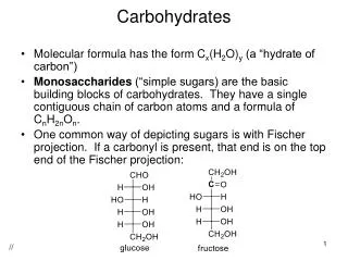
Methods of Glucose Measurement • Glucose has the ability to function as a reducing agent and aid in the detection and quantitation of carbohydrates • Glucose and other carbohydrates are capable of converting copper(II) in an alkaline solution to form copper(I). • The solution loses its deep-blue color and a red precipitate of cuprous oxide forms. • Benedict's and Fehling's reagents, which contain an alkaline solution of cupric ions stabilized by citrate or tartrate, respectively, have been used to detect reducing agents in urine and other body fluids. M. Zaharna Clin. Chem. 2009
Methods of Glucose Measurement • Glucose oxidase method: converts beta-glucose to gluconic acid. • Mutarotase may be added to facilitate the conversion of alpha-glucose to beta-glucose. • Oxygen is consumed and hydrogen peroxide is produced. • Horseradish perixidase is used as a catalyst. Chromagens used for color change M. Zaharna Clin. Chem. 2009
Methods of Glucose Measurement • Hexokinase: more accurate less interference from uric acid, bilirubin and ascorbic acid. • In the presence of ATP- hexokinase converts glucose to glucose-6-phosphate. • Glucose-6-phophate and NADP converted to 6-phosphogluconate and NADPH by glucose-6-phosphate dehydrogenase- absorbance is measured at 340 nm. M. Zaharna Clin. Chem. 2009
Methods of Glucose Measurement M. Zaharna Clin. Chem. 2009
Oral Glucose Tolerance Test (OGTT) • The OGTT continues to be regarded as the most robust means for establishing the diagnosis of diabetes in equivocal cases. • The WHO suggests that only when an OGTT cannot be performed should the diagnosis rely on FPG. • OGTTs should be carried out under controlled conditions after an overnight fast. M. Zaharna Clin. Chem. 2009
OGTT • Collect blood sample while fasting. • The patient is given 75 gm of glucose orally. • Blood samples collected at 60, 120, and 180 minutes. • Analyze the samples and draw a chart. • In normal persons, a return to the fasting level occurs in 2 or at most 2½ h. • In diabetics, the peak is higher and there is a delay in the return of the blood glucose to a fasting level. • Urine remains free from glucose throughout the test in normal individuals and becomes positive in about 60 minutes in diabetics. M. Zaharna Clin. Chem. 2009
OGTT Graph M. Zaharna Clin. Chem. 2009
Tests of Diabetes Control and Disease Progression • Laboratory testing for diabetes after diagnosis of the disease is directed toward the assessment of the progression of disease. • The laboratory offers analysis that helps the physician determine the extent of glycemic control and the risk for the severe consequences of hyperglycemia. M. Zaharna Clin. Chem. 2009
Laboratory tests • Glucose • Glycosylated Hemoglobulin (HbA1c) • Ketone Bodies • Serum osmolality • Electrolytes • Microalbuminuria M. Zaharna Clin. Chem. 2009
Glycosylated Hemoglobulin (HbA1c) • Is a term used to describe the formation of Hb compound when glucose reacts with the amino group of Hb. • Used to monitor and manage diabetes, monitors blood glucose levels over the last 60-90 days. • Specimen of choice is EDTA whole blood M. Zaharna Clin. Chem. 2009
Ketone Bodies • Ketone bodies are produced by the liver through the metabolism of fatty acids to provide energy to provide ready energy from stored lipids • Acetone, Beta-hydroxybutyrate and acetoacetic acid • Low levels present all the time, but when the body is deprived of CHO , ketones levels increase. • Need fresh serum or urine – tightly stoppered and analyzed immediately M. Zaharna Clin. Chem. 2009
Microalbuminuria • Because Diabetes mellitus cause progressive disease in the kidneys (nephropathy), • An early sign that nephropathy is occurring is an increase in urinary albumin • The lab will monitor urinary albumin through measuring microalbumin in the urine. • An annual assessment of kidney function by the determination of urinary albumin excretion is recommended for diabetic patients M. Zaharna Clin. Chem. 2009
Self-Monitoring Glucose Meters • At-home or near-patient monitoring by point of care testing (POCT) with glucose meters provides information so that therapeutic intervention may be initiated immediately. • Glucose meters use the same chemical reactions that are used in glucose analysis in the laboratory: glucose oxidase, hexokinase, and dehydrogenase. • Most systems use dehydrated reagents embedded in pads on plastic strips. M. Zaharna Clin. Chem. 2009
Self-Monitoring Glucose Meters • The strip is inserted in the meter, where the reaction is measured. • The reaction may be a color change that is measured by: • reflectance spectrophotometry, • or the reaction may produce a change in current that can be measured by electrochemistry. M. Zaharna Clin. Chem. 2009
Self-Monitoring Glucose Meters • A blood sample is applied to the surface layer, which both acts as a spreading layer and is a semi-permeable membrane that separates blood cells from plasma. • Plasma from the sample diffuses into the paper analytical layer, which contains the buffered enzyme reaction system, activated by plasma water. • Within the analytical layer, glucose and atmospheric oxygen are acted on by the glucose oxidase to produce hydrogen peroxide and gluconic acid. • In the presence of peroxidase, also contained within the analytical layer, hydrogen peroxide oxidizes a redox indicator to produce a visible colour change. M. Zaharna Clin. Chem. 2009