Download
1 / 24
250 likes | 516 Views
The Physics of Seeing Inside People An introduction to the science of Medical Imaging or “MRI for beginners”. Dr. S. J. Doran. Summary of today’s lecture. What is Medical Imaging? How does MRI work? What can we do with the images?.

E N D
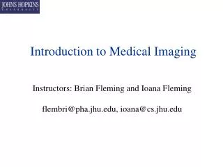
The Physics of Seeing Inside People An introduction to the science of Medical Imaging or “MRI for beginners” Dr. S. J. Doran
Summary of today’s lecture • What is Medical Imaging? • How does MRI work? • What can we do with the images? • How does all this relate to what a typical Physics undergraduate might be doing for the three years at University?
What is Medical Imaging? Hi-tech scanner Images (preferably wacky colours) come out Patient goes in Medical imaging as seen on TV !
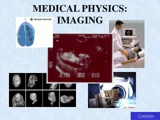
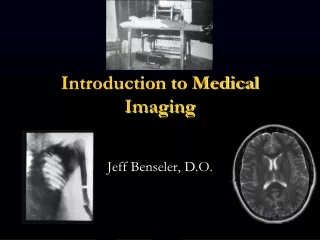
Ultrasound Composite MRI + PET X-ray CT What is Medical Imaging? • The application of basic Physics to see inside the human body • Not one subject but many — lots of different techniques • Each one measures a different physical property of the sample.
Plane-film X-ray maps the total attenuation of X-rays along a path through the body, giving a projection image. Good for bone structure in accidents. Data source : Mayo Clinic • X-ray CT measures the X-ray attenuation coefficient of the body at each point. True 3-D images. Data source : Toshiba America Medical Systems Visualisation : Vitrea 2, Vital Images • Ultrasound maps the reflectionand attenuation of sound. Why use different methods of imaging ? Different methods reveal different features
MRI maps the distribution and “environment” of water molecules in the body. Data source: FORENAP, Rouffach, France • PET maps the distribution of radioactively labelled compounds. • MEG maps directly the magnetic fields generated by currents flowing in the brain. Data source : CSUA, Berkeley Why use different methods of imaging ? Different methods reveal different features (cont.) Data source : SMIS Ltd
Nuclear Physics • Electromagnetism • Mathematics, Signal and Image Processing The Physics of MRI • Classical and Quantum Mechanics • Statistical Mechanics
I B = m0 n I n turns / unit length Magnet for Whole-Body Imager • What is the tunnel into which the patient slides? Image source : GE Medical Systems, VA Imaging Centre, University of Florida
Spin • Nuclear Physics • Many nuclei have an intrinsic spin • A spinning charged particle has a magnetic moment. What happens in a scan? Fundamentals of MRI • Mechanics • Newton’s Law says that a torque will cause the dipole to precess in a direction perpendicular to the torque and the spin vector.
Magnetism • We exert a couple on the nuclear spin by placing a magnetic field across the sample. B m • If the particle were not spinning, it would align like a compass, but, because of the spin, it precesses instead. Precession Spin • Resonance • An electromagnetic field that oscillates at exactly the same frequency as the nucleus will be absorbed. • Under certain conditions, the nucleus will also emit a quantum of RF energy What happens in a scan? Magnetic fields and resonance
B Head Feet x What was happens in a scan? Magnetic field gradients • Resonant frequency is related to the magnetic field. • f B • If we vary the magnetic field across the sample, then the frequency of emitted radio waves varies.
An aerial to transmit and receive radiofrequency signals Image source : GE Medical Systems What happens in a scan? Equipment being used • A large (normally superconducting) magnet • A “gradient set” to create a varying magnetic field • A computer to process the data
Maxwell Pair Saddle Coil / Golay Pair What happens in a scan? The MRI gradient set • Basic Level 2 Physics tells us how to create our magnetic field gradient. • It also explains why the gradients make a knocking noise.
What happens in a scan? Processing the data a Our NMR signal is made up of the sum of lots of different frequencies, corresponding to different spatial positions. b Acquiring multiple signals gives us a 2-D dataset. c Fourier transforms, from Level 2 Maths, can unscramble the data and make an image.
The Human Brain as seen by MRI Data sources : Left - The Whole-brain Atlas, K. A. Johnson and J. A. Becker, Harvard; Right - SMIS UK Ltd.
As it reaches the brain, the signal intensity rises. • We can use this to find out various parameters of the blood supply to the brain tissue. Data source: James D’Arcy and David Collins, Institute of Cancer Research, Sutton Image contrast in the brain (1) • We can inject a chemical called a contrast agent into the bloodstream. • This allows us to calculate the concentration of the agent.
MR images can be sensitised to the rate of diffusion of water molecules. • This allows us to map the local direction of a fibre and create a map of the fibres. • Finally, we can overlay them on a computer model of the head. Data source: Geoff Parker, Institute of Neurology, London Image contrast in the brain (2): neural fibre tracking • Water diffuses faster along nerve fibres than perpendicular to them.
Image contrast in the brain (3): Functional imaging • We acquire one image of the brain in a “resting” state. • We follow this by a corresponding image where the brain is active. • Any differences between these two images correspond to places where the brain is working. Data source: Functional Imaging Laboratory, London Combined PET / MRI study • We can see you think!!
Liver motion during normal breathing Liver Lung Data source : ICR, Sutton Knee sports injuries Lumbar spine Data source : SMIS Data source : SMIS MRI can image much more than just the brain ...
Original MR images Data source : Dr David Lomas, Dept. of Radiology, Addenbrokes Hospital, Cambridge A virtual tour round the human colon ... Computer-generated “flythrough” model
In both cases, one can obtain images of the body’s blood vessels with exquisite detail. More about image contrast … MR angiography • The contrast in MR images can be made sensitive either to “flowing” material, or to an externally administered contrast agent. Prof. Arlart,Katharinen Hosp., Stuttgart, GermanyData source, via GE publicity material
High resolution anatomical imaging • Imaging of coronary arteries MRI in cardiology • “Movies” — data acquired in < 20 s
And finally … the state of the art in cardiac imaging Data source: GE Medical Systems
Conclusion • There are many different ways of imaging the human body. • The different methods tell us different things. • It is study of basic Physics (electromagnetism, nuclear physics, mechanics) which has discovered the principles. • It is money — the human brain is a very valuable thing — which has led to the incredible developments that we see today.