Download
1 / 83
830 likes | 841 Views
This textbook chapter provides an overview of the muscular system, including the types of muscles, their functions, and the structure of skeletal muscles. It also covers common muscle injuries and how muscles contract.

E N D
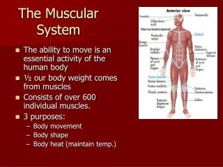
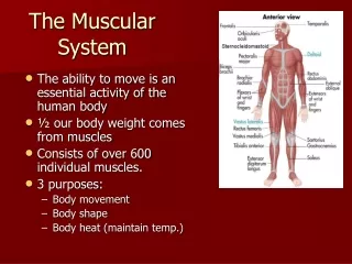
Textbook Chapter 8 The Muscular System
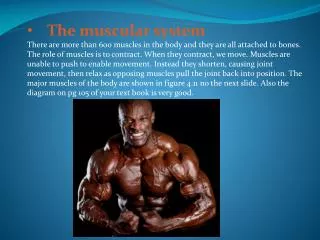
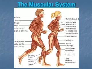
The Muscular System • Muscles are organs composed of specialized cells that use chemical energy, obtained from nutrients, to pull on structures to which they are attached • There are three types of muscles: skeletal, smooth, and cardiac muscle • We will focus on skeletal muscles
The Muscular System • Muscles are responsible for all types of body movement—they contract, or shorten, to make the body move
Functions of Muscles • Support the body • Allow for movement by making bones and other body parts move • Maintain constant body temperature • Assist in movement of cardiovascular veins and lymph vessels • Protect internal organs and stabilize joints
Body System Connections • Integumentary System • The skin increases heat loss during skeletal muscle activity • Lymphatic System • Muscle action pumps lymph through the lymphatic vessels
Structure of a Skeletal Muscle • A skeletal muscle is an organ of the muscular system that is composed of skeletal muscle tissue, nervous tissue, blood, and other connective tissue
Structure of a Skeletal Muscle:Connective Tissue Coverings • Layers of connective tissue enclose and separate all parts of a skeletal muscle • Fascia (dense connective tissue) separates all individual muscles from each other
Structure of a Skeletal Muscle:Connective Tissue Coverings • Epimysium is a layer of connective tissue that closely surrounds each skeletal muscle • Perimysium separate the muscle tissue into small sections called fascicles • Fascicles are bundles of skeletal muscle fibers • Endomysium is a thin covering of connective tissue around a muscle fiber
Skeletal Muscle Attachments • Epimysium blends into a connective tissue attachment • Tendon- cord-like structure • Sites of muscle attachment • Bones • Cartilages • Connective tissue coverings
The Muscular System:Tendonitis • In tendonitis, a tendon becomes painfully inflamed and swollen following injury or repeated stress of athletic activity • The tendons most commonly affected are those associated with the joint capsules of the shoulder, elbow, and hip and those that move the hand, thigh, and foot
Structure of a Skeletal Muscle:Skeletal Muscle Fibers • A skeletal muscle fiber is a single cell that contracts in response to stimulation and then relaxes when the stimulation ends • Skeletal muscle fibers contain many threadlike myofibrils that lie parallel to one another
Structure of a Skeletal Muscle:Skeletal Muscle Fibers • Myofibrils play an important role in muscle contraction • They contain two kinds of protein filaments (myofilaments): • Thin filaments called actin • Thick filaments called myosin • The organization of the myofilaments is what produces alternating light and dark bands of a skeletal muscle fiber
Structure of a Skeletal Muscle:Skeletal Muscle Fibers • The bands of skeletal muscle result from a repeating pattern of units called sarcomeres • The striation pattern has 2 main parts: • I bands (light bands) that are connected to structures called Z lines • A bands (dark bands) which extend the length of the thick filaments
Organization of Skeletal Muscle Fascicle: a bundle of muscle fibers Sarcomere: units of myofibrils responsible for the striated appearance Muscle belly Muscle fiber: muscle cell Myosin: thick filaments Myofilament: protein filaments that make up a sacromere Myofibrils: structures that make up a muscle fiber Actin: thin filaments
Structure of a Skeletal Muscle:Muscle Strains • Muscle fibers and their associated connective tissues are flexible but can tear if overstretched • This is called a muscle strain • The severity of the injury depends on the degree of damage • Mild strains—only a few muscle fibers are injured and fascia remains intact. Loss of function is minimal • Sever strains—many muscle fibers and fascia tear. Muscle function may be completely lost. Painful and produces discoloration and swelling
Structure of a Skeletal Muscle:Neuromuscular Junction • Remember that neurons paly a role in communication within the body • Motor neurons are neurons that control the muscles in the body • Muscle fibers contract when they receive a stimulus from a motor neuron
Structure of a Skeletal Muscle:Neuromuscular Junction • Each skeletal muscle is connected to the end of a motor neuron • The neurons communicate with the muscle fibers through neurotransmitters (chemicals released by the nervous system) • The connection between a motor neuron and the muscle fiber it controls is called a neuromuscular junction
Skeletal Muscle Contraction • A muscle fiber contraction occurs when myosin binds to actin and exerts a pulling force • The result is a movement in which the actin and myosin slide past one another, increasing the area of overlap • This shortens the muscle fiber, which then pulls on the body part that it moves
Skeletal Muscle Contractions • Myosin contains “heads” projecting from one end • Actin contains binding sites for the myosin heads to attach • Actin is often shaped like a double helix—proteins troponin and tropomyosin are part of this helix • A myosin head can attach to a binding site on actin and pull • The myosin then releases the actin and combines with a binding site further down the actin filament
Skeletal Muscle Contraction • The sliding filament model of muscle contraction is based on these actin-myosin interactions • The filaments do not change length, they slide past one another, with the thin filaments moving toward the center of the sarcomere • Acetylcholine serves as the stimulation needed for muscle contraction to begin • Once acetylcholine breaks down and the stimulus to the muscle fiber ends, the muscle relaxes
Body System Connections • Skeletal System • Bones provide attachments that allow skeletal muscles to cause movement • Digestive System • Skeletal muscles are important in swallowing. The digestive system absorbs the nutrients needed for muscle contraction
Energy Sources for Contraction • ATP molecules supply the energy for muscle contraction • When contraction starts, a muscle fiber has a limited amount of ATP • When the fiber is active it must make more ATP in order to keep contracting • A molecule called creatine phosphate contains high energy phosphate bonds that are needed to make ATP • When the supply is exhausted by the muscle fiber, it must rely on cellular respiration to make ATP
Skeletal Muscle Contraction:Steps of Muscle Contraction • Impulse travels down a motor neuron • The neuron releases Acetylcholine (ACh) • ACh binds to receptors in the muscle fiber • An impulse travels over the surface of the muscle fiber • Calcium channels open • Calcium binds to troponin (actin) • Tropomyosin moves to expose binding sites • Myosin heads attach to the actin filaments • Actin is pulled toward the center of the sarcomere • The muscle fiber produces a pulling force
Skeletal Muscle Contraction:Muscle Fatigue • Fatigue is when muscles that have been exercised strenuously for a prolonged period may have decreased ability to contract • Occasionally, muscles become fatigued and cramps at the same time • A cramp is a painful condition in which a muscle undergoes a sustained involuntary contraction • Occurs when extracellular fluid somehow trigger uncontrolled stimulation of the muscle
Muscular Responses • One way to observe muscle contraction is to remove a single muscle fiber from a skeletal muscle in the lab • Threshold Stimulus • Certain strength of stimulation applied to the muscle fiber to begin contraction
Muscular Responses:Recording of a Muscle Contraction • The response of a single muscle fiber to a single impulse is called a twitch • A twitch consists of a period of contraction and a period of relaxation. These can be recorded in a myogram • The twitch has a brief delay between the time of stimulation and the beginning of contraction (latent period) • 2 milliseconds in humans
Muscular Responses:Recording of a Muscle Contraction • The various movements we need to preform daily activities requires contraction of multiple muscle fibers simultaneously • Muscle fibers vary in contraction speed • Fatigue-resistant slow twitch • Fatigueable fast twitch
Muscular Responses:Recording of a Muscular Contraction • Contractions of whole muscles enable everyday activities, but the force generated by contractions must be controlled • EX. Holding a cup • The degree of tension reflects: • The frequency at which the muscle fibers are stimulated • How many fibers take part in the overall contraction
Muscular Responses:Summation • When a muscle fiber is exposed to a series of stimuli of increasing frequency, it is unable to relax between twitches • When the force of the individual twitches combines, it is called summation
Muscular Responses:Recruitment of Motor Units • The higher the intensity of stimulation, the more motor units are activated • An increase in the number of motor units being activated during a contraction is called recruitment • As the intensity increases, recruitment continues until all motor units are activated and the muscle contracts with maximum tension
Musclar Responses: Sustained Contractions • Summation and recruitment together can produce a sustained contraction • Sustained contractions allow us to perform everyday activities • Even when a muscle appears to be at rest, its fibers undergo some sustained contraction--muscle tone • A response to nervous stimulation that originates from the spinal cord and stimulates only a few muscle fibers at a time • Muscle tone is important for posture • If muscle tone is lost, the body collapses
Smooth Muscle • The contraction of smooth muscle is very similar to the contraction of skeletal muscle • There are some important structural and functional differences between smooth and skeletal muscles
Smooth Muscle:Smooth Muscle Cells • Smooth muscles are elongated with tapered ends • Also contain thin and thick filaments, but they are randomly organized • Not striated
Smooth Muscle:Smooth Muscle Cells • There are 2 kinds of smooth muscle cells: • Multiunit smooth muscle • Visceral smooth muscle
Smooth Muscle:Smooth Muscle Cells • In multiunit smooth muscle, the cells are separate from one another • Found in the irises of the eyes and in the walls of blood vessels • Typically contract only in response to stimulation by neurons or certain hormones
Smooth Muscle:Smooth Muscle Cells • Visceral smooth muscle is made up of sheets of cells in close contact with one another • This is the more common type of smooth muscle • Found in the walls of hollow organs (like the stomach, intestines, bladder)
Smooth Muscle:Smooth Muscle Cells • Visceral smooth muscle displays rhythmicity—a pattern of repeated contractions • Due to self-exciting cells that deliver spontaneous impulses periodically into the surrounding tissue • When one cell is stimulated, it causes stimulation in surrounding cells • Ripple effect
Smooth Muscle:Smooth Muscle Cells • Rhythmicity and transmission of impulses are responsible for the wave-like motion (peristalsis) • Peristalsis helps force the contents of certain organs along their length (i.e. the intestines)
Smooth Muscle:Smooth Muscle Contraction • Smooth muscle is slower to contract than skeletal muscle • Can maintain a forceful contraction longer than skeletal muscle • Smooth muscle can stretch as organs fill, yet maintain a constant pressure inside the organs
Cardiac Muscle • Cardiac muscle is found only in the heart • Contraction is the same as skeletal and smooth • There are some important differences
Cardiac Muscle • Cardiac muscle is composed of branching, striated cells • Has many filaments of actin and myosin • Each end of a cardiac muscle cell is connected to an intercalated disc • These allow impulses to pass freely so they travel rapidly from cell to cell • When one part of the network is stimulated, the impulse passes to the rest of the network • The whole structure contracts as a unit
Cardiac Muscle • Cardiac muscle is also self-exciting and rhythmic • A pattern of contraction and relaxation repeats, causing the pattern of the heartbeat
Muscles of Mastication:Masseter • Origin • Zygomatic Arch • Insertion • Posterior lateral surface of the mandible • Action • Elevates and protracts the mandible
Muscles of Mastication:Temporalis • Origin • Temporal bone • Insertion • Mandible • Action • Elevates and retracts the mandible