Download
1 / 48
550 likes | 617 Views
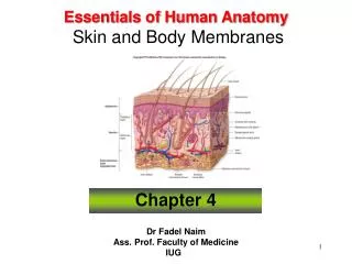
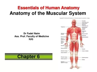
Essentials of Human Anatomy Skin. 1. Skin (integument) is body’s largest organ Approximately 1.6 to 1.9 m 2 in average-sized adult Integumentary system describes the skin and its appendages—the hair, nails, and skin glands Thin and thick skin

E N D
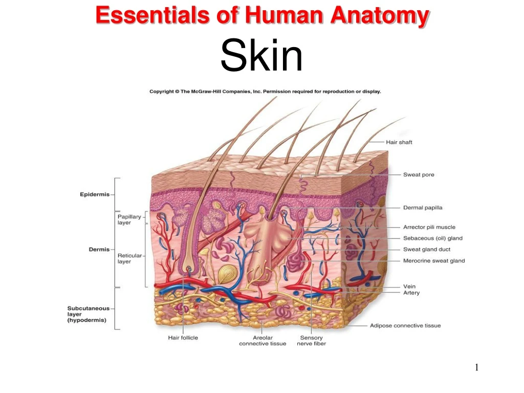
Skin (integument) is body’s largest organ Approximately 1.6 to 1.9 m2 in average-sized adult Integumentary system describes the skin and its appendages—the hair, nails, and skin glands Thin and thick skin “Thin skin”—covers most of body surface (1 to 3 mm thick) “Thick skin”—soles and palms (4 to 5 mm thick) Anatomy of Skin Slide 2
Functions of Skin Function of Integumentary System A. Regulation of Body Temperature B. Protection from Dehydration and Infection C. Respond to Temperature, Pressure, Pain D. Excretion of Water, Salts, Urea (nitrogenous waste) E. Synthesis Vitamin D (essential for Ca + P absorption) F. First Defensive Barrier of Immune Response
Functions of the Skin Protection Physical barrier to microorganisms Barrier to chemical hazards Reduces potential for mechanical trauma Prevents dehydration Protects (via melanin) excess UV exposure Slide 4
Functions of the Skin Sensation Skin acts as a sophisticated sense organ Somatic sensory receptors detect stimuli that permit us to detect pressure, touch, temperature, pain, and other general sensations Slide 5
Functions of the Skin Flexibility Skin is supple and elastic, thus permitting change in body contours without injury Excretion Water Urea/ammonia/uric acid Slide 6
Functions of the Skin Hormone (Vitamin D) production Exposure of skin to UV light converts 7-dehydrocholesterol to cholecalciferol— a precursor to vitamin D Blood transports precursor to liver and kidneys, where vitamin D is produced Process and end result fulfill the necessary steps required for vitamin D to be classified as a hormone Slide 7
Functions of the Skin Immunity Phagocytic cells destroy bacteria Langerhans cells trigger helpful immune reaction working with “helper T cells” Slide 8
Functions of the Skin Heat loss approximately 80% of heat loss occurs through the skin; remaining 20% occurs through the mucosa of the respiratory, digestive, and urinary tracts Slide 9
Layers of Skin • Epidermis • Dermis • Subcutaneous layer • beneath dermis • not part of skin
Epidermis • Lacks blood vessels • Keratinized • Thickest on palms and soles (0.8-1.4mm) • Melanocytes provide melanin • Rests on basement membrane • Stratified squamous
Structure of the Skin Epidermis Cell types Keratinocytes— constitute over 90% of cells present; principal structural element of the outer skin Melanocytes— pigment-producing cells (5% of the total); contribute to skin color; filter ultraviolet light Langerhans cells—dendritic (branched) antigen-presenting cells (APCs), they play a role in immune response Slide 13
Epidermis • Layers of Epidermis • stratum corneum • stratum lucidum • stratum granulosum • stratum spinosum • stratum basale
Structure of the Skin Epidermis Cell layers Stratum germinativum (growth layer)— describes the stratum spinosum and stratum basale together Stratum basale (base layer)—single layer of columnar cells; only these cells undergo mitosis, then migrate through the other layers until they are shed Stratum spinosum (spiny layer)—cells arranged in 8 to 10 layers with desmosomes that pull cells into spiny shapes; cells rich in RNA Slide 15
Structure of the Skin Cell layers Stratum granulosum (granular layer)—cells arranged in two to four layers and filled with keratohyalin granules; contain high levels of lysosomal enzymes Stratum lucidum (clear layer)—cells filled with keratin precursor called eleidin; absent in thin skin Stratum corneum (horny layer)—most superficial layer; dead cells filled with keratin (barrier area) Slide 16
Structure of the Skin Dermal-epidermal junction A definite basement membrane, specialized fibrous elements, and a polysaccharide gel serve to “glue” the epidermis to the dermis below The junction serves as a partial barrier to the passage of some cells and large molecules Slide 17
Structure of the Skin Dermis Sometimes called “true skin”—much thicker than the epidermis and lies beneath it Gives strength to the skin Serves as a reservoir area for storage of water and electrolytes Slide 18
Dermis • On average 1.0-2.0mm thick • Contains dermal papillae • Binds epidermis to underlying tissues • Irregular dense connective tissue • Muscle cells • Nerve cell processes • Specialized sensory receptors • Blood vessels • Hair follicles • Glands
Structure of the Skin Dermis Contains various structures: Arrector pili muscles and hair follicles Sensory receptors Sweat and sebaceous glands Blood vessels Rich vascular supply plays a critical role in temperature regulation Slide 20
Structure of the Skin Dermis Layers of dermis: Papillary layer— composed of dermal papillae that project into the epidermis; contains fine collagenous and elastic fibers; contains the dermal-epidermal junction; forms a unique pattern that gives individual fingerprints Reticular layer— contains dense, interlacing white collagenous fibers and elastic fibers to make the skin tough yet stretchable; when processed from animal skin, produces leather Slide 21
Lines of Cleavage • Tension lines in the skin identify the predominant orientation of collagen fiber bundles. • Clinically and surgically significant because cuts can result in slow healing and increased scarring.
Subcutaneous Layer • hypodermis • loose connective tissue • adipose tissue • insulates • major blood vessels
Structure of the Skin Hypodermis Also called subcutaneous layer or superficial fascia Deep to the dermis, forming connection between the skin and other structures Not part of the skin Slide 26
Skin Color • Genetic Factors • varying amounts of melanin • varying size of melanin granules • albinos lack melanin • Physiological Factors • dilation of dermal blood vessels • constriction of dermal blood vessels • accumulation of carotene • jaundice • Environmental Factors • sunlight • UV light from sunlamps • X rays • darkens melanin
Skin Color Melanin Basic determinant of skin color is quantity, type, and distribution of melanin Beta carotene (group of yellowish pigments from food) can also contribute to skin color Hemoglobin color changes also occur as a result of changes in blood flow Redder skin color when blood flow to skin increases Cyanosis—bluish color caused by darkening of hemoglobin when it loses oxygen and gains carbon dioxide (Figure 6-9) Bruising can cause a rainbow of different colors to appear in the skin Other pigments from cosmetics, tattoos, and bile pigments in jaundice Slide 29
Basis of Skin Color The color of skin and mucous membranes can provide clues for diagnosing certain problems, such as Jaundice yellowish color to skin and whites of eyes buildup of yellow bilirubin in blood from liver disease Cyanosis bluish color to nail beds and skin hemoglobin depleted of oxygen looks purple-blue Erythema redness of skin due to enlargement of capillaries in dermis during inflammation, infection, allergy or burns Slide 30
Sebaceous glands Secrete sebum—oily substance that keeps hair and skin soft and pliant; prevents excessive water loss from the skin usually associated with hair follicles Lipid components have antifungal activity Simple, branched glands Found in dermis except in palms and soles Secretion increases in adolescence; may lead to formation of pimples and blackheads Skin glands Slide 31
Sweat Glands • Widespread in skin • Originates in deeper dermis • Or hypodermis • Eccrine glands • Apocrine glands • Ceruminous glands • Mammary glands
Eccrine glands Most numerous sweat glands; quite small Distributed over total body surface with exception of a few small areas Simple, coiled, tubular glands Function throughout life Secrete perspiration or sweat; eliminate wastes; and help maintain a constant core temperature Sweat glands Slide 33
Apocrine glands Located deep in subcutaneous layer Limited distribution—axilla, areola of breast, and around anus Large (often more than 5 mm in diameter) Simple, branched, tubular glands Begin to function at puberty Secretion shows cyclic changes in female with menstrual cycle Sweat glands Slide 34
Hair Distribution—over entire body except palms of hands and soles of feet and a few other small areas Fine and soft hair coat present before birth called lanugo Coarse pubic and axillary hair that develops at puberty called terminal hair Slide 35
Functions of Hair • Protection • Heat retention • Prevents the loss of conducted heat from the scalp to the surrounding air • Facial expression • Sensory reception • Visual identification • Chemical signal dispersal
Hair Follicles • Epidermal cells • Tube-like depression • Extends into dermis • Hair root • Hair shaft • Hair papilla • Dead epidermal cells • Melanin • Arrector pili muscle
A. Shaft - projects above surface of epidermis 1. medulla - polyhedral cells with eleidin 2. cortex - elongated cells with/out pigment 3. cuticle - outermost layer, like shingles on roof B. Root - below epidermis, penetrates into the dermis C. Hair Follicle - at the base a single hair • external root sheath - basale and spinosum extension • internal root sheath - internal hair cell layers • bulb - base of hair cell • papilla - in the bulb, provides nourishment for hair • matrix - origin of new hair cells D. arrector pili - smooth muscle, cause hair to rise nerve bundle responds to touch E. hair root plexuses
Color result of different amounts, distribution, types of melanin in cortex of hair Growth hair growth and rest periods alternate; hair on head averages 5 inches of growth per year Sebaceous glands attach to and secrete sebum (skin oil) into follicle Male pattern baldness results from combination of genetic tendency and male sex hormones Appearance of hair Slide 42
Hair Thinning and Baldness • Alopecia – hair thinning in both sexes • True, or frank, baldness • Genetically determined and sex-influenced condition
Consist of epidermal cells converted to hard keratin Nail body—visible part of each nail Root—part of nail in groove hidden by fold of skin, the cuticle Lunula—moon-shaped white area nearest root Nails Slide 44
Nail bed—layer of epithelium under nail body contains abundant blood vessels Appears pink under translucent nails Growth—nails grow by mitosis of cells in stratum germinativum beneath the lunula; average growth about 0.5 mm per week, or slightly over 1 inch per year Slide 45
Types of Burns First-degree only epidermis (sunburn) Slide 46
Types of Burns Second-degree burn destroys entire epidermis & part of dermis fluid-filled blisters separate epidermis & dermis epidermal derivatives are not damaged heals without grafting in 3 to 4 weeks & may scar Slide 47
Types of Burns Third-degree or full-thickness destroy epidermis, dermis & epidermal derivatives damaged area is numb due to loss of sensory nerves Slide 48