Download
1 / 22
220 likes | 383 Views
Organ:- Lung Lesions:- Most of the pleural surface covered by abundant fibrin and fibrous tissue DD:- CBPP. Organ:- Chest cavity Lesions:- Large sheets of fibrin cover the costal and diaphragmatic pleura, and form pockets containing straw-colored fluid DD:- CBPP.

E N D
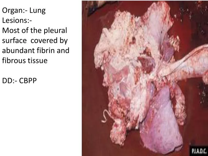
Organ:- LungLesions:-Most of the pleural surface covered by abundant fibrin and fibrous tissueDD:- CBPP
Organ:- Chest cavity Lesions:- Large sheets of fibrin cover the costal and diaphragmatic pleura, and form pockets containing straw-colored fluid DD:- CBPP
Organ:- Costal ribsLesions:-There was a thick plaque (adhesion) of fibrous tissue on the costal pleura.DD:- CBPP
Organ:- LungLesions:-Interlobular septa are markedly thickened by fibrous tissue, and also contain small depressions (air pockets = emphysema). Lobules are reddened and wet (congestionand edema)DD:- CBPP
Organ:- Heart Lesions:-The pericardial sac contains abundant turbid fluid DD:- CBPP
Organ:- Bovine carpus Lesions:- There was abundant fibrin within the synovial space and on the synovium, and articular cartilages contain a few small erosions. DD:- CBPP
Organ:- Lung Lesions:- Presence of Fibrinous exudates in the pleura. DD:- CCPP
Organ:- Carpal joint Lesions:- Haemorrhage on the synovial membrane with periarticular edema in the carpal joint. DD:- Contagious agalactia
Case History • This cow was suddenly found dead with: • Unclotted bloody discharge from all body orifices,. • Incomplete rigor mortis. • Rapid distension of the carcass with gases. • DD:- Peracute anthrax
Case History • This zebra was found dead (after 3 days of suffering from a rapid disease onset) with: • Unclotted bloody discharge from all body orifices. • Incomplete rigor mortis. • DD:-Acute anthrax
Horse Lesions:- Presence of cutaneous nodules (white) on the hind legs. DD:- Glanders
Organ:- Lung Lesions:- Presence of firm, rounded miliary nodules on the lung paranchyma. DD:- Glnaders
Organ:- Equine head Lesions:- Mucopurulent discharge came out from both nostriles DD:- Strangles.
Organ:- Head of equine Lesions:- Hot painful abscesses of the lymph nodes of the throat. DD:- Strangles (Distemper).
Organ:- Equine head Lesions:- Sub-maxillary lymph nodes appear enlarged with present of yellowish creamy Pus. DD:- Strangles
Organ:- Lung Lesions:- Multiple pyo- granulomas are found in the lungs. DD:- Rhodococcus equi infection
Organ:- Kidney of foal Lesions:- (A): The indistinct white foci in the cortex of the kidney are areas of acute infection and contain the bacterium.(B): Infiltration of nuetrophils within glomeruli and tubules and obliterated glomeruli by presence of basaphilic structure ( bacterial colonies). DD:- Septicemia due to Actinobacillus equuli.
Case history:- An affected horse moves with a stiff-legged gait, often with the tail held out stiffly and the ears pricked. As the disease progresses the muscles become very rigid and stiff that the horse may fall and not be able to get up again. DD:- Tetanus
Organ:- Eye of foal Lesions:- The prominence of the foal's third eyelid. DD:- Tetanus
Organ:- Small intestine of pig Lesions:- The mucosa was congested and covered by a large yellowish- brown casts of fibrino- necrotic exudate (diphtheritic pseudomembrane). DD:- Salmonellosis
Case history:- A 48-year-old man presents with draining sinus tracts on his elbow (arrows). The drainage resembles small grains of sand. DD:- Botryomycosis
Organ Skin biopsy obtained from human with hard fibrous nodules on his arm. Lesions:- 1- Basophilic concretion surrounded by an eosinophilic rim is seen 2- The concretion consists centrally of basophilic granular material (bacterial colonies) and peripherally of a homogenous eosinophilic material oriented in radiating club-shaped bodies (rosettes-shaped) about the bacterial colonies. DD:- Botryomycosis