Download
1 / 48
530 likes | 888 Views
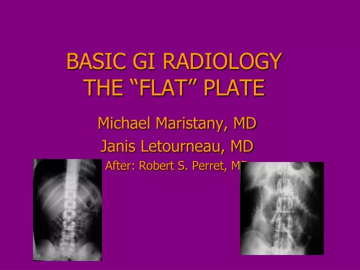
BASIC GI RADIOLOGY THE “FLAT” PLATE. Michael Maristany, MD Janis Letourneau, MD After: Robert S. Perret , MD. KUB/Abdominal plain film. Most common abdominal radiograph Patient supine Often combined with upright (CXR). KUB. Normal KUB. KUB. K kidney U ureter B bladder

E N D
BASIC GI RADIOLOGYTHE “FLAT” PLATE Michael Maristany, MD Janis Letourneau, MD After: Robert S. Perret, MD
KUB/Abdominal plain film • Most common abdominal radiograph • Patient supine • Often combined with upright (CXR)
Normal KUB
KUB • K kidney • U ureter • B bladder • Term KUB indicates that the kidneys, ureters, and bladder are on the film • But these organs are not necessarily seen on the image
K K u u KUB – Backwards u u B
KUB • Paradoxically, organ system of greatest interest is often GI tract • Small bowel situated centrally • Large bowel located on periphery
colon small bowel colon rectum
Contrast filled stomach and small bowel
Contrast filled Colon
Miller-Abbott decompression tube For intestinal obstruction
KUB’sWhy order them, anyway? • > 85% (for pain, colic, nausea, etc) • KUB will be non-contributory or normal
Why can KUB be useful? • Bowel gas pattern characterization • Detection of free air • Abnormal calcifications • Detection of organomegaly • Discovery of abdominal masses • Evaluation of bony structures • Surgical / other medically relevant history
Bowel Gas Pattern • Four major patterns • Ileus • Obstruction • Gasless • Normal • “Free” air
Abnormal Bowel Gas Pattern:Small Bowel Obstruction • Bowel Obstruction • Dilated loops of bowel • SBO – (small bowel obstruction) • Adhesions • Less likely inflammatory/neoplastic • Colonic obstruction • More often of malignant etiology
Small Bowel Obstruction • Dilated loops of small bowel (>3 cm) KUB • With air/fluid levels on upright view • Stair-step pattern to air/fluid levels • Gasless colon
Normal Abnormal
Dilated small bowel - KUB Air/fluid levels on upright
PFs => SBO CT ABDOMEN: SBO Transition point – luminal caliber
Normal KUB
What’s likely dx? Common etiologies?
FREE (INTRAPERITONEAL) AIR KUB is not the best exam; upright or LLQ views helpful Think also of CT; not only good for detection, but for cause
KUB – Abnormal Ca++ • Calcifications • Gallstones • Kidney stones • Vascular • Masses with calcifications (myoma, AAA) • Gallstones • 20% < will be calcified • Kidney stones • >75% will be calcified
KUB - Porcelain Gallbladder (or very large calcified stones)
ERCP Gallstones? Most gallstones will not be seen on KUB – not sufficiently calcified
KUB - kidney stones • Kidney stones will often be visualized • Related to extent of calcification • Detection limit 1-2 mm • Overlying intestinal gas limiting • Obesity limiting
Other Calcified Abnormalities • Uterine myomas • Pancreatic ductal calcifications • Vascular calcification • Appendicolith • Neoplasms • sarcoma, testicular cancer, neuroblastoma • Old hematomas
Calcified Uterine arteries
Splenic hematoma Injection granulomata
Patient Detained Miami International
Non-calcified mass or mass effect • Major limitation of plain films • Organomegaly (multifocal dz vs diffuse) • Neoplasm, abscess, hematoma • Free peritoneal fluid (distribution of SB) • Difficult to differentiate • Relatively homogeneous • Soft tissue density • Merit of CT and MRI
OTHER GIIMAGING MODALITIES • Esophagram • Upper GI Series • Small Bowel Follow-Through • Barium Enema (or Contrast Enema) • CT and CT Colonography • ERCP • MRCP • US
UNKNOWN CASE 52 yo man from Boston Bloody diarrhea Following half-marathon (CCC and poor training)
Thumb-printing: colonic wall edema Inflammation, ischemia, diffuse mural infiltration
CONSIDERATIONSUNKNOWN (REAL) CASES • Patient age and gender • Clinical symptoms • Underlying diseases • Including psychiatric (case of gym sock) • Need for additional views (one at least) • Localize mass or foreign body • Deductive reasoning……….