Download
1 / 16
160 likes | 174 Views
Crystal-storing histiocytosis: definite diagnostic difficulties associated with this marvelous masquerader masking malignant myeloma. Case #: EAHP16-BMWS-224 Annika Windon MD, Yusong Yang MD PhD, Leonas Bekeris MD, Adam Bagg MD. Clinical History. 57-year-old male

E N D
Crystal-storing histiocytosis: definite diagnostic difficulties associated with this marvelous masquerader masking malignant myeloma Case #: EAHP16-BMWS-224 Annika Windon MD, Yusong Yang MD PhD, Leonas BekerisMD, Adam Bagg MD
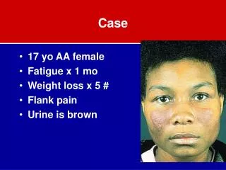
Clinical History • 57-year-old male • Presented with a creatinine of 14 mg/dL in acute renal failure during an episode of severe diarrhea secondary to Clostridium difficile • Bone marrow showed peculiar inclusions in macrophages that were interpreted to be concerning for leishmaniasis slides submitted in consultation
Bone marrow aspirate smear, modified Wright-Giemsa (20x): Expanded macrophage population with intracytoplasmic inclusions
Bone marrow aspirate smear, modified Wright-Giemsa (40x): Macrophages contain abundant irregular purple intracytoplasmic inclusions
Bone marrow aspirate smear, modified Wright-Giemsa (100x): Plasma cells contain similar irregular pinkish-purple intracytoplasmic inclusions
Aspirate clot, H&E (20x): Cluster of macrophages with eosinophilic intracytoplasmic inclusions
Aspirate clot, H&E (50x): Clusters of macrophages with intracytoplasmic crystals
Aspirate clot, H&E (100x): Macrophages and plasma cells with intracytoplasmic crystals
Immunohistochemistry summary • Plasma cell expansion (20-30%) • CD79a+ CD138+ MUM1+ kappa-restricted • CD20- CD45- CD56- PAX5- • Macrophages • CD68+ CD163+ lysozyme+ • Crystalline material within macrophages • Kappa+ CD138+
Aspirate clot, immunohistochemistry, lighter image (50x): CD138 highlights plasma cells and inclusions in macrophages Plasma cells: Macrophages:
Aspirate clot, immunohistochemistry, darker image (40x): CD138 highlights plasma cells and inclusions in macrophages Plasma cells: Macrophages:
Aspirate clot, immunohistochemistry (50x): Plasma cells (brighter) and intracellular inclusions (dimmer) are kappa restricted Kappa Lambda
Cytogenetics • Conventional cytogenetics • Normal male karyotype • FISH • Positive for t(11;14)/CCND1-IGH
Diagnosis • Crystal-storing histiocytosis
Interesting Features • Diagnostic challenges • Macrophages harboring crystalline material often mistaken for Gaucher cells or pseudo-Gaucher cells of CML • This case was thought to reflect leishmaniasis • The macrophages masked the underlying plasma cell neoplasm • Crystals are CD138+ • Raises the possibility that the macrophages may be ingesting plasma cell fragments rather the protein • Prognosis • Typically poor, but this patient remains alive and well 4 years after presentation