Download
1 / 32
320 likes | 443 Views
AD-4 Status Report 2009. 35 Scientists from 12 Institutions. University of Aarhus University Hospital of Aarhus University of New Mexico, Albuquerque University of Athens Queen’s University Belfast Vinca Institute for Nuclear Physics, Belgrade CERN, Geneva Hôpital Universitaire de Geneve

E N D
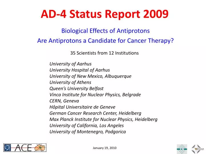
AD-4 Status Report 2009 35 Scientists from 12 Institutions University of Aarhus University Hospital of Aarhus University of New Mexico, Albuquerque University of Athens Queen’s University Belfast Vinca Institute for Nuclear Physics, Belgrade CERN, Geneva Hôpital Universitaire de Geneve German Cancer Research Center, Heidelberg Max Planck Institute for Nuclear Physics, Heidelberg University of California, Los Angeles University of Montenegro, Podgorica Biological Effects of Antiprotons Are Antiprotons a Candidate for Cancer Therapy?
Rationale for Conformal Radiotherapy Dose (and tumor control) are limited due to tolerance of organs at risk Dose to Target Better conformity of dose to target enables application of higher doses & higher tumor control without increasing normal tissue complication rate
Particle Therapy offersReduced Integral Dose to Body Particles deposit LESS physical dose in front of the tumor and NO dose beyond the distal edge of the Bragg peak!
Monte Carlo Comparisons for different Particles MCNPX FLUKA GEANT SHIELD HIT . . Beware of GIGO! We need Experiments
The AD-4 Experiment at CERN • INGREDIENTS: • V-79 Chinese Hamster cells embedded in gelatin • Antiproton beam from AD(126 MeV) • METHOD: • Irradiate cells with dose levels to give survival in the peak is between 0 and 90 % • Slice samples, dissolve gel, incubate cells, and look for number of colonies ANALYSIS: • Study cell survival in peak (tumor) and plateau (skin) and compare the results to protons (and carbon ions)
Biological Analysis Method Co-60 Dose RBEplat = Plateau Dose =1.0 RBEpeak = Co-60 Dose =1.15 Peak Dose • Plot “peak” and “plateau” survival vs. relative dose to extract the Biological Effective Dose Ratio BEDR = F • RBEpeak/RBEplateau (F = ratio of physical dose in peak and plateau region) Plot “peak” or “plateau” survival vs. absolute dose and compare to 60Co irradiation comparing dose values needed for Iso-Effect for peak, plateau, and 60Co irradiation: Relative Biological effectiveness RBE
Carbon Ions – SOBP at GSI note: clinical beams with precise dosimetry and fast dose delivery …….. Higher energy to achieve clinical relevant depth and form SOBP….
RBE for Carbon Ions Extract survival vs. dose plot for each depth slice and calculate RBESF=10% RBEplateau = 1.2 RBEpeak = 2.0 RBE distal = 1.5
CERN DATA 2006 ANTIPROTONS note: insufficient data due to dose error (beam spot size) …….
CERN DATA 2007 note: poor quality of SOBP due to degrading mechanism …….
CERN DATA 2008 note: better control over dose planning for SOBP……..RBE = 1.2 RBE = 1.73 – 2.2 RBE = 1.52 (???)
CERN DATA 2009 note: attempt to collect data in tail for RBE analysis……..
Combining/Comparing Data ? Different ratios of low LET/high LET in SOBP inhibit combination of data except for plateau
RBE for Antiprotons(Plateau) RBEplat = 1.25 Good agreement with Monte Carlo Results (Bassler, Holzscheiter; Acta Oncologica, 2009; 48: 223 226)
RBE for Antiprotons(Peak) RBEplat = 1.25 RBEPeak = 2.25 Need more data points for fit. Need repeat experiments for statistics. But everything points towards “step-function” for RBE vs. depth for antiprotons
Potential Clinical Advantages? • Each Particle Type shows distinct features • Protons are well known and easy to plan (RBE = 1) which is the reason they are most widely adopted. • Antiprotons have lowest entrance dose for the price of an extended isotropic low dose halo. • Carbon ions have sharpest lateral penumbra but comparatively higher entrance dose than even protons (no RBE included here), but show forward directed tail due to in beam fragmentation. Detailed dose plans (including RBE) will need to be developed to assess applicability of particle types for different tumor types and locations!
How to Tell the Difference? Dose Volume Histograms (DVH’s) show what percentage of the volume of interest receiveswhat level of dose. These are the standard figures of merit for dose planners. two opposing fields Antiprotons produce less radiation load to healthy tissue than protons or carbon ions !!!(carbon ions and antiprotons considered biological identical) Single field
DNA Damage Assays • There is more to biology than just clonogenics – especially outside the targeted area: • Immediately after attack on DNA proteins are recruited to the site • This event signals cell cycle arrest to allow repair • If damage is too extensive to repair programmed cell death (apoptosis) is induced • Cells also deficient of cell cycle check point proteins may enter mitosis (cancer cells are often deficient in repair proteins and continue dividing) g-H2AX: Phosphorylation of H2AX in the presence of Double Strand Breaks Micronuclei: Fluorescent detection of micronuclei (parts of whole chromosomes) formed due to DNA damage indicating potential of tumorigenesis g-H2AX and Micronucleus assays are typically used to study immediate and long term DNA damage respectively
Liquid Ionization Chambers • Higher density of liquid medium than air provides: • Higher sensitivity • Allows smaller dimensions • Measurement of steep field gradients • Useable at very low dose rate Tetramethylsilane (TMS) • Higher density means higher recombination losses • General recombination • Ions generated by different incident particles recombine • Dose rate dependence • Initial (columnar) recombination • Ions created by same incident particle recombine • depends on ionization density along track • can be used to determine Linear Energy Transfer (LET) C8H18 Isooctane • Columnar recombination is described by Jaffe Theory • Understand relationship of columnar and general recombination ongoing experiments at AD-4 and Umeo Microtron
LIC Results Measure charge collected for different voltagesin Plateauin Bragg Peak 2 mm and 5 mm distal to Bragg Peak Asymptotic slope is indicationof Linear Energy Transfer Jaffe Theory can be usedto calculate flux averaged LET Results This work: LETantiprotonBP = 9.0 keV/mm LETproton(173 MeV)BP = 3.2 keV/mm Kempe et al. LETproton(202 MeV)BP = 3.6 keV/mm
New Beam Monitor • Purpose: Shot-to-shot monitoring of beam spot shape, size, and position • to replace Gafchromic film, facilitate alignment, and have instant feed back on beam changes • Solution: Solid state pixel detector (Monolithic Active Pixel Sensor) • Dedicated MAPS design to beam monitoring • pixel 153×153µm2 squares • two 9×9 interdigited arrays of n-well/p-epi diodes + two independent read-out electronics – avoiding dead time • In-pixel storage capacitors – choice ~0.5pF or ~5pF to cope with signal range Mimotera, Massimo Caccia (Universita’ dell’Insumbria Como, Italy) Long term goal: Measure LET distributions in 2D/3D
First Results – All Single Shots Single shot pixel matrix x – y slices through several shots to check stability of beam • Initial test of Mimotera in AD-4 (2 hours of beam time) • Joint effort to quantify response to intensity and LET while using Mimotera as online beam monitor • Additional tests and R&D at HIT to certify detector for medical use Lego representation of beam profile
Real Time Imaging - Simulation • Use 4 x 3 layers of (virtual) silicon pixel detectors 40x40 cm2 / 5 cm spacing 30 cm from origin • Pixel Resolution s = 100 mm • Track charged pions and photons • Overall detector efficiency e = 1% • Define vertex as approach of two tracks closer than 2 mm • If more than 2 tracks per particle are detected use meta-center of vertices of any two tracks
Results of Simulation Beam Eye View Side View Achievable Precision: +/- 1mm (Accepted for publication in Phys. Med. Biol.)
First Experimental Realization • Q: How to minimize material and cost • A: Instead of 3 layers use one layer and look at grazing incidence. • Q: What detector to use for first test?A: Turn to our friends in ALICE and use one (spare) module of the Alice Silicon Pixel Detector (SPD)
First Experimental Realization pbar Water phantom • grazing incidence of pions produce long tracks • length distribution changes with angle • stopping distribution along z-axis can be inferred • Future work: Expansion to 3 - D 289.5° Bragg Peak 293.0° distal fall-off π±
Summary and Outlook Achievements • Initial Results on Biological Effect of Antiprotons can be used for preliminary Dose Planning Studies • RBE Enhancement confined to Bragg Peak only • DNA damage assays for studies of late effects • Initial Steps towards Fast Beam Monitoring and Real Time Imaging of Stopping Distribution • Initial Progress towards LET Measurement All this with 6 weeks of beam time (1.2 x 10¹² pbars)
Summary and Outlook Recent Publications • J.N. Kavanagh, F.J. Currell, D.J. Timson, M.H. Holzscheiter, N. Bassler, R. Herrmann, G. Schettino; ‘Induction of DNA Damage by Antiprotons for a Novel Radiotherapy Approach’; submitted to European Physical Journal D (2009) • Niels Bassler, Ioannis Kantemiris, Julia Engelke, Michael Holzscheiter, Jørgen B. Petersen, ‘Comparison of Optimized Single and Multifield Irradiation Plans of Antiproton, Proton and Carbon Ion Beams’, submitted to Radiotherapy and Oncology (2009) • Bassler, N., Holzscheiter, M.H., Petersen, J.B., ‘Neutron Fluence in Antiproton Radiotherapy, Measurements and Simulations', submitted to Physics in Medicine and Biology (2009) • Kantemiris, I., Angelopoulos, A., Bassler, N., Giokaris, N., Holzscheiter, M., Karaiskos, P., Kalogeropoulos, T.E., ‘Real-time imaging for dose evaluation during antiproton irradiation’, Physics in Medicine and Biology, in print (2009) • Kovacevic, S., Bassler, N., Hartley, O., Vranjes, S., Garaj-Vrhovac, V., Holzscheiter, M., ‘V-79 Chinese Hamster Cells irradiated with antiprotons, a study of peripheral damage due to medium and long range components of the annihilation radiation’, Int. J. of Rad. Biology 85: pp. 1148-1156 (2009) • Fahimian, B.P., DeMarco, J.J., Keyes, R., Bassler, N., Iwamoto, K.S., Zankl, M., Holzscheiter, M.H., ‘Antiproton radiotherapy: peripheral dose from secondary neutrons’, Hyperfine Interaction 194: pp. 313-318 (2009) • Bassler, N., Holzscheiter, M. 2009, ‘Calculated LET Spectrum from Antiproton Beams Stopping in Water’, Acta Oncologica 48: pp. 223-226 (2009)
Summary and Outlook Future Work • Increase precision of RBE determinationBetter biological protocolsHigher data density to stabilize fits • Continue DNA damage studies to assess risk of secondary primary malignancies (SPM’s) • Detailed dose planning studies on specific cancers • Develop 2D LET measuring system • Complete real-time imaging pilot experiment • Transfer technology between HIT and AD-4 - Protons, Carbon ions, Antiprotons
Beam Time Request 1 week of 126 MeV (500 MeV/c) antiprotons • Survival measurements on V-79 cell lines • Study of DNA damage using H2AX and MN • Liquid ionization chamber measurements • “Standard” 5 MeV (100 MeV/c) antiprotons • Commissioning of modified beam line • Real time imaging • Beam monitor development • Response of detectors and Gafchromic film to annihilation events