Download
1 / 26
420 likes | 899 Views
Histology of cartilage. Introduction. Modified (solid type) of connective tissue. Forms the skeletal basis of some parts of the body. Avascular E.g. Auricle of the ear, lower part of the nose. Components. Cells. Matrix. Chondrocytes. Fibers Collagen OR Elastic fibers

E N D
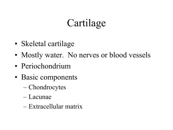
Introduction • Modified (solid type) of connectivetissue. • Forms the skeletal basis of some parts of the body. • Avascular • E.g. Auricle of the ear, lower part of the nose. Histology of cartilage
Components Cells Matrix Chondrocytes • Fibers • Collagen OR • Elastic fibers • Ground substance • Tissue fluid • GAG & proteoglycans Histology of cartilage
Chondrocytes • Derived - chondroblast • Chondroblast - mesenchymal cell • Seen in spaces- lacunae Histology of cartilage
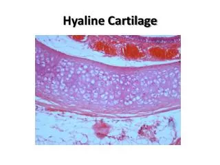
3 types of cartilage • Hyaline cartilage • Elastic cartilage • Fibrocartilage Histology of cartilage
Matrix (fibers + ground substances) • Type II collagen fibers • Proteoglycans, Hyaluronic acid • Capsule or territorial matrix • Interterritorial matrix • Lacunae(tiny spaces in the matrix) • Isogenous cell nest / CELL NESTS (offspring of a common parent cell). Histology of cartilage
Seen under microscope Histology of cartilage
Hyaline cartilage Chondrogenic layer Fibrous layer Perichondrium Chondroblasts Cell nest Matrix Chondrocyte in lacuna
Hyaline cartilage- magnified Terretorial matrix Interterretorial matrix Chondrocyte in lacuna Cell nest
Distribution • Costal cartilage • Articular cartilage • Thyroid, cricoid, arytenoid • Tracheal rings • Part of nasal septum • Epiphyseal plate Histology of cartilage
Elastic cartilage (yellow fibrocartilage) • Greater flexibility. • Ground substance contains abundance of branching and anastomosing elastic fibres. • No isogenous cell nests. • Chondrocytes are found in single or in small groups. Histology of cartilage
Elastic cartilage Histology of cartilage
Elastic cartilage (microscopic view) Histology of cartilage
Elastic cartilage Fibrous layer Chondrogenic layer Perichondrium Chondroblast Matrix with elastic fibers Chondrocyte in the lacuna
Distribution • Auricle & lateral part of the external acoustic meatus. • Medial part of auditory tube. Histology of cartilage
Epiglottis, corniculate, cuneiform, apical part of arytenoid. Histology of cartilage
White fibrocartilage • It is intermediate between Dense connective tissue and hyaline cartilage (tensile strength) • Chondrocytes are found in singles or in rows with bundles of collagen with little matrix. • No perichondrium. • Bundles of collagen (type1) Histology of cartilage
White fibrocartilage Histology of cartilage
White fibrocartilage (microscopic view) Histology of cartilage
White-fibro cartilage Chondrocyte Matrix
Distribution • Secondary cartilaginous joints • intervertebral discs, • pubic symphysis. Histology of cartilage
Articular discs Histology of cartilage
Menisci of the knee joint Histology of cartilage
Growth of Cartilage • Interstitial Growth ?? • Appositional Growth?? Histology of cartilage