Download

1 / 30
300 likes | 321 Views
PROTEIN MODEL CHALLENGE 2019. Lin Wozniewski lwoz@iun.edu. Disclaimer. This presentation was prepared using draft rules. There may be some changes in the final copy of the rules. The rules which will be in your Coaches Manual and Student Manuals will be the official rules.

E N D
PROTEIN MODELCHALLENGE2019 Lin Wozniewski lwoz@iun.edu
Disclaimer This presentation was prepared using draft rules. There may be some changes in the final copy of the rules. The rules which will be in your Coaches Manual and Student Manuals will be the official rules
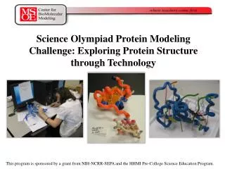
What is Protein Modeling? • Protein modeling involves: • Building a protein model (using toobers or other materials) • Guided by visualization (using Jmol/JSmol) • Of 3D coordinates (available from RCSB PDB) • To understand the structural basis of protein stability, interactions and functions • What will students learn? • The shape, assembly and interactions of proteins • Understand chemical principles of stability and function in these molecules • Apply chemical principles to design new molecules.
Parts of Protein Modeling-The Competition • Pre-build model • Students will bring their pre-built models to the assigned impound. These can be made of toobers or ANY other material • On-site build model • Students will sit at a computer and build a specific region of a protein with the toober provided, guided by visualization of 3-D coordinates from the RCSB PDB, using Jmol/JSmol program • Exam • Students will answer questions on a test • The test will cover structure and properties of amino acids, levels of protein structure (primary, secondary, tertiary, quaternary), important interactions contributing to the function of the protein being studied.
What do the Students need to bring? • For impound • The Pre-build model including suitable additions highlighting functionally significant regions (max=2’X2’X2’) with scale 1 residue = 2 cm • A filled out form indicating what has been added, what it represents and why it is important. This needs to be on a 4”X6” notecard in 3 columns appropriately labeled. • For on-site build • Ability to visualize and explore specified regions of a given protein structures using Jmol/Jsmol • Ability to make models using the provided toobers and side chains • A ruler and a marker • For the exam • Something to write with • Up to 1 page (8.5 X 11”) with appropriate notes and references
What will MSOE and RCSB PDB provide? • For the Pre-build • Pre-build scoring rubrics • 3-D printed model (judge’s model) for event supervisors to use and keep • For the On-site build • On-site build toobers • A CD with Jmol/JSmol and all files needed for visualization of the specified region of the protein • On-site build scoring rubrics • 3-D printed model (judge’s model) for event supervisors to use and keep • For the Exam • The test • Answers to the test
What the event supervisor needs to provide • Enough expertise to be able to interpret and use the provided rubrics to • judge the models and • grade the exam. • Computers for each participating team with • Jmol/JSmol program installed and • All necessary files for the event copied to the computer.
What the students need to do to Prepare • Get familiar with protein structure. Learn about • Levels of protein structure - primary, secondary, and tertiary and quaternary structure • Properties of amino acid side chains – which ones are acid vs basic and hydrophobic vs hydrophilic etc. • Interactions in protein structure – covalent (peptide bonds and disulfide bridges) and non-covalent (hydrogen bonds, hydrophobic interactions, charge based interactions), metal coordination. • Learn to visualize and explore protein structures. • Learn about the Protein Data Bank (www.rcsb.org) • Learn to use Jmol/JSmol to visualize structures from the PDB
What the students need to do to Prepare • Practice protein modeling • Mark the toober (or other similar material) in 2 cm segments. Each of these segments denote one amino acids in the protein. This corresponds to the primary structure. • Visualize the different alpha helix and beta strand elements in Jmol/JSmol and fold corresponding regions of the toober or other material accordingly. This is the secondary structure. • Fold all secondary structural elements with respect to each other so that they come together to form the 3-D shape of the protein. This is the tertiary structure of the region being modeled. • Identify functionally important structural features to answer questions and/or add creative enhancements to your model. (see following slide).
What the students need to do to Prepare • How to determine the functionally important structural features? • Read the manuscript describing the structure, review article or Molecule of the Month feature provided. Pay attention to any specific amino acid residues, secondary structural elements etc. that are discussed in the context of the protein function. • Visualize the full molecule at the RCSB PDB. Pay close attention to all polymer chains in the structure and how they interact with each other. Usually amino acid residues at the interaction interface have important roles in the function of the protein. Also pay attention to chemical principles in protein folding and stability – hydrophobic amino acids in the core of globular structures, polar and charged amino acids on the surface etc.
Background info about 20 amino acids • Backbone consist of: • Amino group (NH2 or NH3+) • Carbon atom, where the side chain is bound • Carboxyl group (COO- or COOH) • Side chains are either: • Hydrophobic • Have only carbon and hydrogen atoms • Usually buried inside the protein • Non-polar or apolar • Hydrophilic • have hydroxyl, carboxylic acid, or amine groups • Are generally on the outside of the protein • May be acidic or basic, or polar
Primary Structure of a Protein The Primary Structure of the Protein is the actual order of the amino acids in the protein. This is the next thing the students must determine after they learn about amino acids & the side chains Lys- Glu-Thr-Arg-Arg-Arg-Lys-etc.
Secondary Structure of a Protein The 2 most common types of secondary structure are the alpha helix and beta pleat The alpha helix ONLY coils right-handed (if you are going up the stairs, your right hand rests on the outside banister going up) The beta pleat should bend back and forth in a zigzag pattern of about 20 at each start of a new amino acid Alpha Helix Beta Pleat
Tertiary Structure of a Protein The tertiary structure of the protein is the final folding that is the result of the molecular interactions formed by the primary and secondary structure. This is determined using the J-Mol program. What a finished pre-build might look like Pipe Cleaner My personal favorite - 12 Gauge Wire Toober
Resources From MSOE From RCSB PDB • Lending library • http://cbm.msoe.edu/teachRes/library/ • Jmol Program • http://cbm.msoe.edu/scienceOlympiad/sampleEnvironment/zincFingerSample.html • Science Olympiad related material • http://cbm.msoe.edu/scienceOlympiad/index.php • PDB-101 • www.rcsb.org/pdb-101/ • Molecule of the Month features • http://www.rcsb.org/pdb/101/motm_archive.do • Exploration of specific PDB entries • www.rcsb.org • Science Olympiad related material • http://education.pdb.org/olympiad/
A mini event • Make a small part of one protein (a zinc finger) using resources that a team will have during an on-site build. • Look at the scoring rubrics for the model and how to use them. • Score your own model. • The Challenge: • Use the 1ZAA pdb file, create an image in Jmol at www.rcsb.org, identify key structural features, and use it to fold a Mini-Toober model • Note: During the actual competition teams will have access to a computer with a specific version of Jmol/JSmol and necessary files. They will not have access to the internet.
Pdb Database • Type 1zaa into PDB look-up window & hit search. • Click on the sequence tab • Complete sequence is there • Click anywhere on sequence-Jmol will come up and show you molecule (or click on display Jmol button) • Scroll down so that you see poymer 3 chain again. Jmol window will come with you. • Put pointer over any residue & leave pointer there for a couple of seconds. The amino acid letters will appear.
Jmol • Right Click and move mouse to move molecule • Main commands • Backbone • Cartoon • Ribbon • Wireframe • Spacefill • Type command and then a number (1-499 work) • Click on Execute • To get rid of that picture type the command and then 0 • Click on Execute • Hold mouse over one spot for a second or two to see letters & position of amino acid
Guide to making a Zinc finger • Visualize the backbone of amino acids 4-31 of the Zif268 protein (PDB ID 1ZAA) and use it to build a model, which has: • Two β strands starting near the amino terminus, making a β sheet • Has one α chain near the carboxylic end • Has one zinc ion at the center of the structure • Two Histidines side chains (25 & 29) and two Cysteine side chains (7 & 12) coordinate the zinc ion and stabilize the structure. • The structure has hydrophobic core with residues such as phenylaninine 16 & leucine 22 • The Arginine 18 side chain interacts with the DNA
Scoring Zinc Finger-Score Sheet Rubric-Sample Regional-
Full Rubric Sheet N-terminus Beta-sheet Alpha-helix C-terminus Whole Molecule Overview
Questions? Thank You
Rubric Details Blue cap on N-terminal amino acid (Pro4) (1 pt) To receive this point, the blue cap should be positioned on the first amino acid. This should be next to the beta-strand. See picture to the right for correct placement of the blue end cap. Red cap on C-terminal amino acid (Gly31) (1 pt) To receive this point, the red cap should be positioned on the last amino acid. This should be next to the alpha-helix. See picture to the right for correct placement of the red end cap. Alpha helix (amino acids 19-31) is located at C-terminus of protein (2 pts) There should be an alpha helix located at the C-terminus of the protein. See figure to right. On the model and in the figure, the alpha helix is colored magenta.
Rubric Details, Continued Alpha helix is right-handed (2 pts) Alpha helices are right-handed. Check the alpha helix in the model to confirm that the helix is right-handed. If the alpha helix is right-handed, the model is awarded two points. To determine if the helix is right-handed, find one of the ends of the helix and imagine that the helix is a spiral staircase. Pretend that you are climbing that staircase and the helix is the hand-rail, which is always on the ourside edge of the staircase. If you would put your right hand on the toober as you go up the staircase, you have a right-handed helix. If you would put your left hand on the toober, you have a left-handed helix and the model would not receive the points. Alpha helix is properly formed (helix resembles a telephone cord) (1 pt) The helix should be formed in such a way that it resembles a telephone cord stretched out slightly. The helix should not be compacted down so that there is not any space between the turns. It should also not be so stretched out that there is a lot of space between the turns Alpha helix is appropriate length (13 amino acids; ~3.5 turns) (2 pts) The helix is 13 amino acids, and each turn in the helix is approximately 3.6 amino acids in length. Therefore, the length of this helix should be ~3.5 turns.
Rubric Details, Continued Beta strand #1 (amino acids 5-7) (2 pts) To receive these points, the model should have a beta strand from amino acids 5-7 (3 amino acids in length). The first beta strand should be located near the blue end cap. The model and the figure to the right have the beta strands colored yellow. . Beta strand #2 (amino acids 14-16) (2 pts) To receive these points, the model should have a beta strand from amino acids 14-16 (3 amino acids in length). The second beta strand is located 6 amino acids (12 cm) away from the first. The model and the figure to the right have the beta strands colored yellow. Beta strand is formed properly (1 pt) To receive this point, the model should have properly formed beta strands. The model can have the beta strands in a zig-zag shape (a bend every 2 cm) or it could have them be represented as straight regions to the model. There should not be any helical or coiled portions in this area
Rubric Details, Continued Helix is arranged next to beta sheet (protein should be compact with a 2-stranded beta sheet lying next to an alpha helix; helix and sheet should not be too far apart) (2 pts) To receive these points, the beta sheet and alpha helix should be located close to one another. There should only be enough space between the two secondary structure to allow for a zinc ion to coordinate between the 4 amino acids that bind the ion. In other words, there should not be much space between the alpha helix and the beta sheet 12. N-terminus (blue cap) and C-terminus (red cap) are pointed in opposite directions (2 pts) To receive these points, the N-terminus and C-terminus of the protein should be facing away from each other. If you hold the model so that the beta sheet is facing the left and the helix is on the right (like the picture shown to the right), then the N-terminus should be pointing upward and the C-terminus is pointing downward. Please note that there is not much space between the two secondary structures. N-terminus C-terminus
Rubric Details, Continued Model should be flat in that the beta strands and alpha helix are occupying the same plane (2 pts) To receive these points, the alpha helix and beta sheet should be in the same plane (please see figure to the right). The model should be “flat” in that neither the helix nor the sheet protrudes upward or downward form the main axis. You should be able to look through the beta sheet and see the alpha helix Creative Additions to model (2 pts each): Zinc ion To receive these points, the model should have a zinc ion located between the alpha helix and beta sheet closer to the C-terminus than the N-terminus. Please model and figure to the right (zinc ion is colored dark red). 2 Histidines (His 25 and 29) (coordinates Zn) 2 Cysteines (Cys 7 and 12) (coordinates Zn) Cysteine To receive these points, the model should have 2 Cysteines at positions #7 and #12. If zinc ion is present, then these Cysteines should be connected to the zinc ion. Arginine 18 (attaches to DNA) Arginine To receive these points, the model should have an Arginine at position 18. If DNA is present on model, this amino acid should interact with the DNA. To receive these points, the model should have 2 Histidines at positions #25 and #29. If zinc ion is present, then these Histidines should be connected to the zinc ion
Phenylalanine Arginine Leucine Rubric Details 6: To receive point(s) … • Creative Additions to model (two points each): • Arginine 18 bound to DNA (2 pts) • The model should have an Arginine at position 18. • If DNA is present on model, this amino acid should interact with the DNA. • Hydrophobic amino acids (Phe16, Leu22) (2 pts) • These residues should face inward to create a stable hydrophobic core stabilizing protein • DNA attached to protein (2 pts) • The model should have DNA bound to the zinc finger. • Zinc finger should be in the major groove of the DNA.
Rubric Details 7: To receive point(s) … • Creative additions are appropriate (2 pts) • The creative additions should be relevant to telling the functional story of the protein. • Any amino acid shown should play an important role in the stability (Zinc ion coordination, hydrophobic core) or function of the molecule (binding to DNA). • Models that have displayed all of the amino acids should not be awarded these points. • Creative additions are accurate (2 pts) • The creative additions need to be accurate and reflect the scientific information that has been provided in Goodsell’s Molecule of the Month, the PDB file or alternative resources. • Students submitted a 3x5 card to explain model (2 pts) • A 3x5 card should be submitted along with the model, describing what additional features have been added to the model and what they represents.