Download
1 / 60
600 likes | 606 Views
IMMUNe system. Functions of immune system Terminologies used Types of immunity Structure of immune system organs & cells HLA antigen/ MHC molecule Transplant rejections. Immunity and immunopathology are proverbial two edges of ‘double-edged sword’. Immunity defence mechanism

E N D
Functions of immune system • Terminologies used • Types of immunity • Structure of immune system organs & cells • HLA antigen/ MHC molecule • Transplant rejections
Immunity and immunopathology are proverbial two edges of ‘double-edged sword’. • Immunity defence mechanism • Injurious to human body
FUNCTIONS OF IMMUNE SYSTEM • Recognition of self from non-self • Mounting a specific response against non-self • Memory of what was earlier recognised as non-self • Antibody formation • Cell mediated reactions
Normal function of immunity is for body defence. • Its failure or derangements results in various diseases 1. Immunodeficiency disorders 2. Hypersensitivity reactions 3. Autoimmune disease 4. Amyloidosis
TERMINOLOGIES USED • Antigen (Ag) is defined as a substance, usually protein in nature, which when introduced into the tissues stimulates antibody production. • Haptenis a non-protein substance which has no antigenic properties, but on combining with a protein can form a new antigen capable of forming antibodies.
Antibody (Ab) is a protein substance produced as a result of antigenic stimulation. • Circulating antibodies are immunoglobulins (Igs) of which there are 5 classes: IgG, IgA, IgM, IgE and IgD. • An antigen may induce specifically sensitised cellshaving the capacity to recognise, react and neutralise the injurious agent or organisms.
Antigen may combine with antibody to form antigen antibody complex. • Reaction of Ag with Ab in vitro may be primary or secondary phenomena. • In vivo, the Ag-Ab reaction may cause tissue damage
Divided into 2 types, each with humoral and cellular components: • Natural or innate immunity • Specific or adaptive immunity
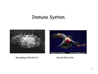
Natural or innate immunity • Non-specific • First line of defense without antigenic specificity. • It has 2 major components: a) Humoral: comprised by complement. b) Cellular: consists of neutrophils, macrophages, and natural killer (NK) cells.
Specific or adaptive immunity • Specific and is characterised by antigenic specificity. • 2 main components: a) Humoral: consisting of antibodies formed by B cells. b) Cellular: mediated by T cells.
ORGANS OF IMMUNE SYSTEM a) Primary lymphoid organs: i) Thymus ii) Bone marrow b) Secondary lymphoid organs: i) Lymph nodes ii) Spleen iii) MALT (Mucosa-Associated Lymphoid Tissue located in the respiratory tract and GIT).
CELLS OF IMMUNE SYSTEM i) Lymphocytes ii) Monocytes and macrophages iii) Mast cells and basophils iv) Neutrophils v) Eosinophils
Lymphocytes • Lymphocyte is the master of human immune system. • Morphologically, lymphocytes appear as a homogeneous group. • Functionally two major lymphocyte populations, T and B lymphocytes are identified. Third type, NK (natural killer) cells.
Formed from lymphoid precursor cells in the bone marrow. • Lymphocytes undergo maturation and differentiation in the bone marrow (B cells) and thymus (T cells) and acquire certain genetic and immune surface characters which determine their type and function; this is based on cluster of differentiation (CD) molecule on their surface. • Identified by ‘CD markers’ by specific monoclonal antibody stain employing immunohistochemistry or by flow cytometry. • B and T lymphocytes proliferate into ‘memory cells’ impart long lasting immunity against specific antigens.
B cells differentiate into plasma cells form specific antibodies • T cells get functionally activated on coming in contact with appropriate antigen. • Upon coming in contact with antigen, it is the macrophage, i.e. specialised antigen-presenting cell such as dendritic cell, and the major histocompatibilty complex (MHC) in the macrophage, which determines whether the invading antigen is to be presented to B cells or T cells. • Strong antigens that cannot be dealt by antibody response from B cells such as certain microorganisms (e.g. viruses, mycobacteriaM. Tuberculosis and M. leprae), cancer cells, tissue transplantation antigen etc are presented to T cells.
B CELLS • Involved in humoral immunity by inciting antibody response. • Comprise about 10-15% of lymphocytes. • On coming in contact with antigen (e.g. invading microorganims) B cells are activated to proliferate and transform into plasmacytoid lymphocytes then into plasma cells. • Common B cell markers include: CD 19, 20, 21, 23. • Also possess B cell receptors (BCR) for surface immunoglobulins (IgM and IgG) and Fc receptor for attaching to antibody molecule. • T cell help is provided to B cells by a subset of T helper cells, TH 2, by elaborated interleukins (IL-4, IL-5, IL-10, IL-13).
T CELLS • Incite cell-mediated immunity and delayed type of hypersensitivity. • Comprise 75-80% of lymphocytes. • Pan T cell markers are CD3, CD7and CD2. • Also carry receptor (TCR) for recognition of MHC molecules. • Depending upon functional activity, T cells have two major subtypes: T helper cells and T suppressor cells.
T helper cells (TH cells) • Promote and enhance the immune reaction and are also termed as T-regulatory cells. • Carry CD4 molecule on their surface and hence are also called CD4+ cells. • CD4+ cells in circulation are about twice the number of CD8+ cells (CD4+/CD8 ratio 2:1). • Act by elaboration of variety of cytokines.
T helper cells.... • Depending upon the type of cytokines elaborated TH cells are further of two subclasses: - TH 1 cells elaborate IL-2 and interferon (IFN)-γ. - TH 2 cells elaborate IL-4, IL-5, IL-6, and IL-10. • CD4+ cells are predominantly involved in cell-mediated reactions to viral infections (e.g. in HIV), tissue transplant reactions and tumour lysis.
T suppressor cells (TS cells) • Suppress immune reactions but are cytotoxic and actually destroy the invading antigen; hence are also termed as cytotoxic T lymphocytes (CTL). • Carry CD8 molecule on their surface and hence are also called CD8+ cells. • CD8+ cells in circulation are about half the number of CD4+ cells. • CD8+ cells are directly cytotoxic to the antigen. • CD8+ cells are particularly involved in destroying cells infected with viruses, foreign cells and tumour cells.
NATURAL KILLER (NK) CELLS • Comprise about 10-15% of circulating lymphocytes. • Do not have B or T cell markers, nor are these cells dependent upon thymus for development. • NK cells carry surface molecules of CD2, CD16 and CD56. • NK cells are large granular lymphocytes. • Part of the natural or innate immunity.
NATURAL KILLER (NK) CELLS... • Recognise antibody-coated target cells and bring about killing of the target directly; this process is termed as antibody-dependent cell-mediated cytotoxicity (ADCC). • Against viruses and tumour cells.
Monocytes and Macrophages • Circulating monocytes are immature macrophages and constitute about 5% of peripheral leucocytes. • Remain in circulation for about 3 days before they enter tissues to become macrophages. • Subpopulations dendritic cells found in the lymphoid tissue and Langerhans’ cells seen in the epidermis, are characterised by the presence of dendriticcytoplasmic processes and are active in the immune system.
Salient features and important immune functions of macrophages are as follows: 1. Antigen recognition 2. Phagocytosis 3. Secretory function 4. Antigen presentation
1. Antigen recognition. • Possess cell surface receptors to several extracellular molecules— receptor for cytokines, component of complement (C3b), selectins, integrins and Fc (constant fragment) of antibody. • Antigen to become recognisable can also get coated by antibodies or complement, the process being termed as opsonisation. • Macrophages have capacity to distinguish self from non-self by presence of human leucocyte antigens (HLA) or major histocompatibilty complex (MHC).
2. Phagocytosis. • Antigen that has been recognised by the macrophages due to availability of surface receptors, or the opsonised antigen, is ready to be engulfed by the process of cell-eating by macrophages.
3. Secretory function. i) Cytokines (IL-1, IL-2, IL-6, 8, IL-10, IL-12, tumour necrosis factor-α) and prostaglandins (PGE, thromboxane-A, leukotrienes) chemical mediators of inflammation and activate other leucocytes. ii) Secretion of proteins involved in wound healing e.g. collagenase, elastase, fibroblast growth factor, angiogenesis factor. iii) Acute phase reactants e.g. fibronectin, microglobulin, complement components.
4. Antigen presentation. • When macrophages are unable to lyse an antigen or an organism act as antigen-presenting cells for presenting to immunocompetent T cells (subtype CD4+ or CD8+ cells), or to B cells. • Lymphoid cell would then deal with such antigen.
Basophils and Mast Cells • Basophils are a type of circulating granulocytes (0-1%) • Mast cells are their counterparts seen in tissues (connective tissue around blood vessels and in submucosal location). • Have IgE surface receptor. • In contact with antigen binding to IgE (e.g. allergic reaction to parasites) cells get activated release granules i.e. degranulate. • Granules contain substances such as: histamine, platelet activating factor, heparin and certain chemical mediators (e.g. prostaglandins, leukotrienes). • Mediate inflammation in allergic reactions and have a role in wound healing.
Neutrophils • Most numerous of the circulating leucocytes (40-75%). • Cytoplasm of PMNs contains lysosomal granules of three types: primary (azurophilic), secondary, and tertiary. • Referred to as ‘microphages’ role as first line of defense against an invading foreign organism in the body. • Act against bacteria and small foreign particulate material.
Eosinophils • Eosinophils are also circulating granulocytes (1-6%). • Role in allergic reactions and in intestinal helminthiasis. • Granules of eosinophils contain lysosomal enzymes, peroxidases, and chemical mediators of inflammation (e.g. prostaglandins, leukotrienes). • On comingin contact with IgE opsonised antigen (e.g. helminths) eosinophilsdegranulate release the chemicals stored in granules incite inflammation.
HLA stands for Human Leucocyte Antigens • These antigens or genetic proteins in the body determine one’s own tissue from non-self (histocompatibility). • First discovered on the surface of leucocytes. • Important in matching donor and recipient for organ transplant, they are called major histocompatibility complex (MHC) or HLA complex.
Most of the transplantation antigens or MHC are located on a portion of chromosome 6 of all nucleated cells of the body and platelets. • Genes occupy four regions or loci—A, B, C and D, on the short (p) arm of chromosome 6 and exhibit marked variation in allelic genes at each locus. • Product of HLA antigens is highly polymorphic. • HLA system is part of immunoglobulin superfamily of CAMs.
Depending upon the characteristics of MHC, they have been divided into 3 classes: • Class I MHC antigens • Have loci as HLA-A, HLA-B andHLA-C. • CD8+ (i.e. T suppressor) lymphocytes carry receptors for class I MHC used to identify class I antigen on them. • Class II MHC antigens • Have single locus as HLA-D. • Have further 3 loci: DR, DQ and DP. • Class II MHC is identified by B cells and CD4+ (i.e. T helper) cells.
Class III MHC antigens • Are some components of thecomplement system (C2 and C4) coded on HLA complex • Not associated with HLA expression and are not used in antigen identification.
ROLE OF HLA COMPLEX 1. Organ transplantation. • Major importance of HLA system is in matching donor and recipient for tissue transplantation. • Recipient’s immune system can recognise the histocompatibility antigens on the donor organ and accordingly accept it or reject it. • Both humoral as well as cell-mediated immune responses are involved in case of genetically non-identical transplants.
2. Regulation of the immune system. • Class I MHC antigens regulate the function of cytotoxicT cells (CD8+ subpopulation) e.g. in virus infections. • Class II MHC antigens regulate the function of helper T cells (CD4+ subpopulation).
3. Association of diseases with HLA. i) Inflammatory disorders e.g. ankylosingspondylitis. ii) Autoimmune disorders e.g. rheumatoid arthritis insulindependent diabetes mellitus. iii) Inherited disorders of metabolism e.g. Idiopathic haemochromatosis.
According to the genetic relationship between donor and recipient, transplantation of tissues is classified into 4 groups: 1. Autografts donor and recipient is the same individual. 2. Isografts donor and recipient of the same genotype. 3. Allografts donor is of the same species but of a different genotype. 4. Xenografts donor is of a different species from that of the recipient.
Skin grafts and organ transplants such as kidney, heart, lungs, liver, pancreas, cornea and bone marrow. • Most commonly practised are skin grafting, and kidney and bone marrow transplantation. • For any successful tissue transplant without immunological rejection, matched major histocompatibility locus antigens (HLA) between the donor and recipient are of paramount importance.
Graft versus- host (GVH) reaction • In humans, when immunocompetent cells are transplanted to an immunodeficient recipient e.g. when severe combined immu- nodeficiency is treated by bone marrow transplantation. • Clinical features include: fever, weight loss, anaemia, dermatitis, diarrhoea, intestinal malabsorption, pneumonia and hepatosplenomegaly. • Intensity of GVH reaction depends upon the extent of genetic disparity between the donor and recipient.