Download
1 / 1
10 likes | 171 Views
Figure 2. XRD patterns of Cloisite 20A and NR/Cloisite 20A nanocomposites. I w. I n. I n / I w. 0,55. 0,24. 0,44. ESR Study of NR/Montmorillonite Nanocomposites M. Didović 1 , D. Klepac 1 S. Valić 1,2 A.P. Meera 3 and S. Thomas 3

E N D
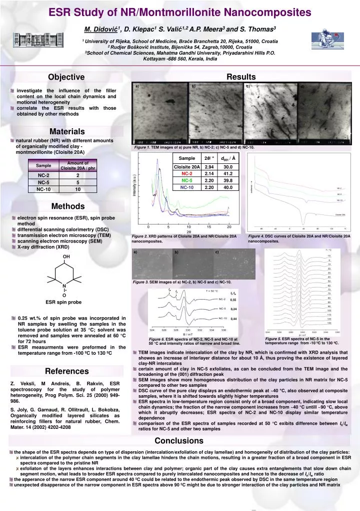
Figure 2. XRD patterns of Cloisite 20A and NR/Cloisite 20A nanocomposites. Iw In In/Iw 0,55 0,24 0,44 ESR Study of NR/Montmorillonite Nanocomposites M. Didović1, D. Klepac1S.Valić1,2 A.P. Meera3 and S. Thomas3 1 University of Rijeka, School of Medicine, Braće Branchetta 20, Rijeka, 51000, Croatia 2 Rudjer Bošković Institute, Bijenička 54, Zagreb,10000, Croatia 3School of Chemical Sciences, Mahatma Gandhi University, Priyadarshini Hills P.O. Kottayam -686 560, Kerala, India Results Objective a) b) c) d) • investigate the influence of the filler content on the local chain dynamics and motional heterogeneity • correlate the ESR results with those obtained by other methods Materials • natural rubber (NR) with different amounts of organically modified clay - montmorillonite(Cloisite 20A) Figure 1. TEM images of a) pure NR, b) NC-2; c) NC-5 and d) NC-10. Methods • electron spin resonance (ESR), spin probe method • differential scanning calorimertry (DSC) • transmission electron microscopy (TEM) • scanning electron microscopy (SEM) • X-ray diffraction (XRD) Figure 4. DSC curves of Cloisite 20A and NR/Cloisite 20A nanocomposites. c) a) b) Figure 3. SEM images of a) NC-2, b) NC-5 and c) NC-10. ESR spin probe • 0.25 wt.% of spin probe was incorporated in NR samples by swelling the samples in the toluene probe solution at 35 °C; solvent was removed and samples were annealed at 60 °C for 72 hours • ESR measurments were preformed in the temperature range from -100 ºC to 130 ºC Figure 5. ESR spectra of NC-5 in the temperature range from -10 ºC to 130 ºC. Figure 6. ESR spectra of NC-2, NC-5 and NC-10 at 50 °C and intensity ratios of narrow and broad line. • TEM images indicate intercalation of the clay by NR, which is confirmed with XRD analysis that showes an increase of interlayer distance for about 10 Å, thus proving the existence of layered clay-NR intercalates • certain amount of clay in NC-5 exfoliates, as can be concluded from the TEM image and the broadening of the (001) diffraction peak • SEM images show more homogeneous distribution of the clay particles in NR matrix for NC-5 compared to other two samples • DSC curve of the pure clay displays an endothermic peak at ~40 °C, also observed at composite samples, where it is shifted towards slightly higher temperatures • ESR spectra in low-temperature region consist only of a broad component, indicating slow local chain dynamics; the fraction of the narrow component increases from ~40 °C untill ~90 °C, above which it abruptly decreases; ESR spectra of NC-2 and NC-10 display similar temperature dependence • comparison of the ESR spectra of samples recorded at 50 °C exibits difference between In/Iw ratios for NC-5 and other two samples References Z. Veksli, M Andreis, B. Rakvin, ESR spectroscopy for the study of polymer heterogeneity, Prog Polym. Sci. 25 (2000) 949-986. S. Joly, G. Garnaud, R. Ollitrault, L. Bokobza, Organically modified layered silicates as reinforcing fillers for natural rubber, Chem. Mater.14 (2002) 4202-4208 Conclusions • the shape of the ESR spectra depends on type of dispersion (intercalation/exfoliation of clay lamellae) and homogeneity of distribution of the clay particles: • intercalation of the polymer chain segments in the clay lamellae hinders the chain motions, resulting in a greater fraction of a broad component in ESR spectra compared to the pristine NR • exfoliaton of the layers enhances interactions between clay and polymer; organic part of the clay causes extra entanglements that slow down chain segment motion, what leads to broader ESR spectra compared to purely intercalated nanocomposites and hence to the decrease of In/Iw ratio • the apperance of the narrow ESR component around 40 ºC could be related to the endothermic peak observed by DSC in the same temperature region • unexpected disapperance of the narrow component in ESR spectra above 90 ºC might be due to stronger interaction of the clay particles and NR matrix