Download
1 / 15
150 likes | 233 Views
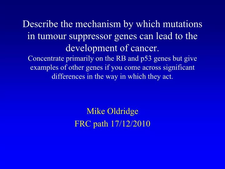
Describe the mechanism by which mutations in tumour suppressor genes can lead to the development of cancer. Concentrate primarily on the RB and p53 genes but give examples of other genes if you come across significant differences in the way in which they act. Mike Oldridge FRC path 17/12/2010.

E N D
Describe the mechanism by which mutations in tumour suppressor genes can lead to the development of cancer. Concentrate primarily on the RB and p53 genes but give examples of other genes if you come across significant differences in the way in which they act. Mike Oldridge FRC path 17/12/2010
Essay plan Intro to cancer – uncontrolled cell division: 6 general features 6 or 7 steps towards cancer - highly unlikely to happen in single cell 2 mechanisms increase likelihood of this progression: growth advantage, genomic instability Oncogenes vs TS genes – differences between 3 mechanisms of TS action: inappropriate cell cycle progression, apoptosis, maintenance of genome stability Classical TS – RB1 Describe retinoblastoma Knudson 2 hit model Describe functions of RB1 – control of G1-S phase progression. Diagram showing interactions P53 Describe function – role in cell cycle progression and apoptosis Predominantly point mutations – gain of function? Li-Fraumeni Protein interactions – diagram Third mechanism – genome stability Chrm level: spindle checkpoint, dna damage, telomeres DNA level: dna repair – other genes involved
Cancer Condition in which cells divide without control Result of somatic cells acquiring genetic changes that confer 6 general features: • Independence of external growth signals • Insensitivity to external anti-growth signals • Ability to avoid apoptosis • Ability to replicate indefinitely • Ability of a mass of such cells to trigger angiogenesis and vascularise • Ability to invade tissues and establish secondary tumours
Cancer These features are acquired by natural selection – each successive mutation confers an extra selective advantage to descendants of a cell Many interlocking and overlapping defence mechanisms: Potential tumour cells are either repaired and brought back into line or made to undergo apoptosis It is thought that six or seven independent defences need to be disabled for a normal cell to be converted into a malignant tumour - highly unlikely that this would happen in a single cell 2 mechanisms exist that allow this progression to happen – an initial mutation can increase the likelihood of subsequent mutations by Conferring a growth advantage – thereby increasing the target population for the next mutation Inducing genomic instability at DNA or chromosomal level – thereby increasing overall mutation rate Takes time to accumulate mutations – generally late onset disease
Oncogenes vs Tumour Suppressor genes Oncogenes Normal activity promotes cell proliferation Complex regulation limits this activity Gain of function mutations: point mutations, copy number amplification, chromosomal rearrangements → excessive or inappropriate activity Tumour Suppressor (TS) genes Normal function limits cell proliferation – 3 mechanisms Prevent inappropriate cell cycle progression Control apoptosis Maintain genome stability and keep mutation rates low by ensuring accurate replication, repair and segregation of cell’s DNA Loss of function mutations to both alleles remove this complex regulation
Classical tumour supressor – RB1 Retinoblastoma – aggressive childhood cancer of the eye Variable phenotype - Unilateral/bilateral tumours 60% are unilateral cases - usually sporadic 40% are bilateral - always heritable although the proband is often the 1st case in the family Only 10% have a family history Transmitted as an incompletely penetrant dominant disorder in 1971 Knudson noted age of onset for bilateral cases was consistent with 1 mutation whereas sporadic cases fitted with 2 hit kinetics
2 hit model Familial retinoblastoma linked to chm 13q14 Cavenee et al (1983) showed loss of one copy of this region in DNA from sporadic tumour patients compared to normal cells from the patients Loss of heterozygosity – many TS genes identified by this method Point mutations Promoter methylation – tumour DNA is hypomethylated but promoters often methylated leading to silencing
RB1 • RB1 encodes 928 aa nuclear phosphoprotein RB • Part of small family of nuclear proteins including p107 and 130 (not thought to be altered in tumours but may provide redundancy) - ‘pocket proteins’ • Share sequence similarity in 2 areas which constitute the A/B pocket and bind E2F family of transcription factors • C-ter contains cyclin-cdk interaction motif, can also bind MDM2 • Also has roles in apoptosis and differentiation Progression through cell cycle controlled by cyclins and cyclin dependent kinases RB has prominent role as ‘gatekeeper’ that negatively regulates progression through G1 phase of cell cycle
RB – cellular function E2F1 transcription factor – 1st cellular target of RB Regulates genes involved in G1-S transition Negatively regulated by RB RB negatively regulated by CDK4/6 – phosphorylation of RB by CDK disrupts RB/E2F association CDK4/6 in turn regulated by p16 (CDKN2A) RB phosphorylated 2-4 hrs before cell enters S phase – allows E2F to activate genes necessary for progression to S phase p16, CDK4/6 and RB have been found to be altered in wide variety of tumours Classon et al
P53 • 53kDa phosphoprotein encode by TP53 • Family includes TP63 and 73 – can also induce apoptosis and share binding sites with P53: overlapping and distinctive functions • Known as ‘Guardian of the genome’ due to central role in preventing inappropriate cell cycling / apoptosis • Cell cycle stalls in cells with damaged DNA – if damage not repairable → apoptosis. Tumour cells with absent/non functional p53 continue to replicate damaged DNA and do not undergo apoptosis • Mutated in about half of cancers resulting in loss of apoptotic function • Unusually, most mutations are point mutations – gain of function? • Constitutional mutations in TP53 cause Li-Fraumeni syndrome – cancer predisposition with high penetrance (90%) of multiple primary tumours
P53 • p53 levels normally low because ubiquitylation by MDM2 targets it for degradation • Signals from stress sensors (DNA damage, hypoxia, heat shock, metabolic changes and certain cytokines) lead to phosphorylation of p53 – no longer a substrate for MDM2 and hence levels of p53 rise • Increases transcription of genes such as: • p21 – inhibitor of Cdk2 and cell cycling • PUMA, BAX, NOXA – control apoptosis
Genome stability Third mechanism by which TS genes act is in stabilising the genome at either the chromosomal or nucleotide level Chromosome instability: Spindle checkpoint: chromatids separate before correctly attached DNA damage: chrm abnormalities can be biproduct of attempts at DNA replication with damaged DNA Telomeres: become too short to protect chrm ends → structural abnormalities DNA level instability: Nucleotide excision repair Base excision repair – MUTYH Double strand break repair – BRCA1/2 Replication error repair: cause generalised increase in mutation rates → microsatellite instability – MLH1/MSH2
References • Classon et al. (2002) Nat Rev Cancer 2: 910-917 • Lohmann DR and Gallie BL (2004) Am J Med Genet, 129C 23-28 • Steele RJ at al (1998) Br J Surg, 85, 1460-67 • Vousden KH and Liu X (2002) Nat Rev Cancer,2, 594-604 • Strachan and Read 4th edition
Key words Cancer Oncogenes vs tumour suppressors Knudson’s 2 hit model Retinoblastoma – RB1 Growth advantage Genomic instability Cell cycle regulation Apoptosis P53