Download
1 / 26
260 likes | 345 Views
Vision: Stimulus and physiology. What is light? One kind of electromagnetic radiation (emr includes lots of other stuff, like radio waves, x-rays, radar waves and so forth) EMR behaves like particles and waves.

E N D
What is light? One kind of electromagnetic radiation (emr includes lots of other stuff, like radio waves, x-rays, radar waves and so forth) EMR behaves like particles and waves. Particle: a particle of light is called a photon; the more photons are being emitted by something, the brighter it is. Light / electromagnetic radiation
EMR behaves like particles and waves (cont.) • Wave: Every kind of light has a specific wavelength; that is, the distance it takes the wave to complete a cycle (start up, come down, then go back up).
What is light? (cont.) • lightis what we call the particular range of emr that we can see. ROY G. BIV, etc.
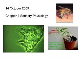
What is light? (cont.) • Other animals can perceive longer or shorter wavelengths. • To you: • To a bee:
Eyeball physiology: beginning the transduction process – from crabs . . .
Eyeball physiology: beginning the transduction process –. . . to vertebrates
Important parts of vertebrate eyes: • lens: used to focus image on back of eye • retina: sensitive to light; transduces energy from light to neural impulses & does preliminary processing • sclera: white part • pupil: black hole • iris: acts like a camera f-stop; lets in the right amount of light for the situation • cornea: first part of light-bending process- to focus image on the eye: two-thirds of bending happens here { contact lens == artificial cornea
Important parts of vertebrate eyes (cont.): • lens: second part of light-bending process; you choose how much to bend the light, to help focus
Important parts of vertebrate eyes (cont.): • Cilliary muscles: control the thickness of the lens
The retina: Light Bipolar cells photoreceptors Ganglion cells
The retina: • Photoreceptors: light-sensitive cells – they send neural signals when light hits them • photopigment: molecule that transforms when light hits it • Rods: contain photopigment rhodopsin (sensitive to a broad range of light) - only allows black and white vision • Cones: contain three different photopigments (each sensitive to a somewhat smaller range of light)
The retina (cont.): • The fovea: small region in the center of the retina. Only contain cones; used for color vision, fine details - vision is sharpest here. • Bipolar cells: Pool information from multiple photoreceptors. • Ganglion cells: Center-surround receptive field; takes input from a number of bipolar cells, some of which activate and some of which inhibit activation of the ganglion cell.
The retina (cont.): • Ganglion Cells (cont.) • Receptive field (cont.)
The retina (cont.): • Ganglion Cells (cont.) • Receptive field (cont.) • Foveal ganglion cells: very small receptive field ≈ 6 bipolar cells • Peripheral ganglion cells: much larger receptive fields
To the brain (cont.)! • Optic nerve: no photoreceptors! ganglion cells bunch together and leave the eye, headed for the brain at this point. One spot in your vision is always blind. • Optic Chiasm: All input from the right visual hemifield goes to the left side of the brain, & vice versa. This means half of the input from the left eye must cross right, etc. This happens at the optic chiasm. • Lateral Geniculate Nucleus: first stop; inputs from eyes (and other areas of brain) • Superior Colliculus: involved in control of eye movements; also receives input from ears & skin.
Visual cortex: • Simple cell:oriented edge detectors (or line detectors); take advantage of center-surround ganglion cell organization to do so.
Visual cortex (cont.) • Orientation tuning curveplots the response of a simple cell across different line orientations • column: vertical series of cells in each layer (i- vi) of primary visual cortex, all responsive to lines of the same orientation. • ocular dominance column: Hubel and Weisel found rows of columns favoring stimulation from either the left or right eye.
Visual cortex (cont.) • retinotopic organization: one column analyzes one point of the visual world imaged on the retina. columns (actually hypercolumns) near one another analyze points near each other in the retina. • end-stopped cellresponds best if the line ends within its receptive field. • Complex cellLarger receptive field; responds best to moving lines, usually in a particular direction. • Feature detectors,Angle-detectors, length detectors, width detectors, pretty much any visual feature - even faces!
Visual cortex (cont.) • Face-detector cell
Visual cortex (cont.) • Hand-detector cell
Visual cortex (cont.) • Grandmother cells? Somewhat tongue-in-cheek term for the idea that there might be cells that activate only when one's grandmother comes into view. • Even celebrities: Quiroga et al. (2005) found single cells in human cortex that respond when shown pictures of Jennifer Aniston, but nothing else. Even similar celebrities like Julia Roberts failed to activate the cell. (So maybe we shouldn't be so tongue-in-cheek about grandmother cells)
Interesting visual disorders: • visual agnosia: inability to identify objects (can still remember, draw, copy) • prosopagnosia: inability to identify faces (even your own!)
List of terms, section 5 • Receptive field • Fovea • Optic nerve • Optic chiasm • Lateral geniculate n ucleus • Superior colliculus • Simple cell • Edge detector • Orientation tuning curve • column/ocular dominance column • End-stopped cell • Complex cell • Retinotopic organization • Feature detector • Grandmother cell • Visual agnogia • prosopagnosia • electromagnetic radiation • Light • Pinhole pupil • Lens • Retina • Sclera • Pupil • Iris • Cornea • Cilliary muscles • Photoreceptors • Photopigment • Rods • Cones • Bipolar cells • Ganglion cells