Download
1 / 16
160 likes | 173 Views
Describe the structure and function of the heart. Explain the role of the heart and blood vessels in the circulatory system. Appreciate the role of science in society and how society influences scientific research. Starter Questions:

E N D
Describe the structure and function of the heart. • Explain the role of the heart and blood vessels in the circulatory system. • Appreciate the role of science in society and how society influences scientific research.
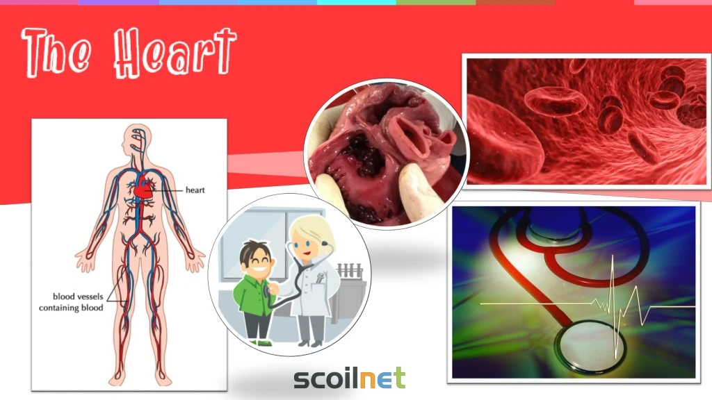
Starter Questions: Using the human model, describe the location of the heart. What protects the heart? If you listen to a heart using a stethoscope, what does it sound like? When does the rhythmic beating of the heart begin? When does the rhythmic beating of the heart stop? What do you think the job of the heart is? Note: If you do not have a human model, you could use this online tool. Click here
Compare 19 grams to 250 grams by weighing out. Your heart pumps blood all over the body. Each person’s heart is a little larger than the size of the person’s fist. A newborn baby’s heart weighs about 19 grams and an adult’s heart weighs about 250-315 grams. The heart is made of a strong cardiac muscle that continuously contracts and relaxes. Every time that your heart contracts, blood is forced out of your heart and into your arteries. This force pushes blood through your arteries in spurts. With each spurt, a beat can be felt. This beat is called a pulse. Model the action of the heart with your fist. Locate the best places to take your pulse. Extend activity with this animation.
Use the videos below as a guide… How fast does your heart beat? To check your pulse at your wrist, place two fingers between the bone and the tendon over your radial artery — which is located on the thumb side of your wrist. When you feel your pulse, count the number of beats in 15 seconds. Multiply this number by four to calculate your beats per minute. Take three measurements and calculate the average. Repeat the procedure after 2 minutes of running on the spot. Extend the activity using this graph. Note: Teachers could project an online timer onto the board. Try this one…
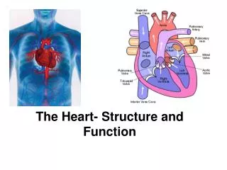
LeftSide Watch the following clip and label the parts of the heart in the given diagram? • Septum • Right Atrium • Left Atrium • Right Ventricle • Left Ventricle • Aorta • Pulmonary Artery • Vena Cava • Pulmonary Vein • Valve RightSide Video Link: http://columbiasurgery.org/heart/about-heart or demonstrate the structure using a dissection of a cow or sheep heart.
The heart is separated into the left side and the right side by a muscular wall called the septum.
There are four chambers in the heart. Left Atrium Right Atrium Pumps blood down to the left ventricle. Pumps blood down to the right ventricle. Left Ventricle Right Ventricle Pumps blood out the Aorta to all around the body. Pumps blood out of the pulmonary artery to the lungs. Draw arrows to direct the blood flow within the heart.
Aorta Blood is pumped away from the heart to all around the body. Blood is oxygenated. Vena Cava Pulmonary Vein Blood enters the heart from the arms, legs and other parts of the body.Blood is deoxygenated. Blood from the lungs is pumped to the heart. Blood is oxygenated. Pulmonary Artery Discuss the location of the values. Blood is pumped Away from the heart to the lungs. Blood is deoxygenated. Recap the function of the valve with this animation.
Colour the deoxygenated blood blue and the oxygenated blood red. Note: Blood vessels transport blood from the heart and to the heart. There are three types of blood vessels… Recap with this animation.
Lungs The steady pumping of the heart supports life by moving blood through the body. Both sides of the heart pump blood at the same time. As the right ventricle contracts and sends blood to the lungs, the left ventricle also contracts and squeezes blood out to the body. Activity – Record the pathway of the blood using the illustration on the right. Body Note: There is a nice activity here on page 30 and 32.
Blood pressure is the force with which the blood pushes against the walls of the arteries as it circulates the body. Blood pressure is measured using a piece of equipment called a sphygmomanometer or digital blood pressure meters Doctors always give blood pressure as two readings. 1. The first number is the higher pressure caused as the left ventricle contracts to push blood out of the heart. This is the systolic blood pressure. 2. The lower value measures the force your heart exerts on the walls of your arteries in between beats. This is the diastolic blood pressure.
Complete research on the variety of diagnostic tools that exist to help diagnose heart conditions. Medicine has made some of its most exciting advances in cardiology. Cardiology is the branch of medicine that deals with diseases of the heart and blood vessels. In the 1900’s, doctors learned to diagnose and treat many heart conditions.
Cardiology Electrocardiogram (ECG): MRI: Doppler Ultrasound: Digital Echocardiography: CAT/CT Scan: MRA:
Cardiology Electrocardiograph: Twelve electrodes are placed on the skin to pick up the electrical pattern of the blood. A record called an electrocardiogram (ECG) is produced, which displays the electrical activity of the heart muscle. (Source: World Book Online) MRI: A diagnostic technique that is used to assess anatomy and heart muscle function, as well as to identify the presence of any scar tissue.(Source: Columbia Surgery) Digital Echocardiography: This technique uses sound waves to produce a video image of your heart. This test can help doctors see the size and shape of your heart along with any abnormalities. It can tell how well your heart is pumping. (Source: Mayo Clinic) Sample Research Coronary Angiography: This technique is usually performed only if diagnosis is difficult. In this test, doctors insert a long, flexible tube called a catheter through a large blood vessel, usually an artery in the area where the thigh and abdomen meet. They push the catheter up to the beginning of the coronary arteries, then inject special dye. The dye shows the condition of the arteries as it travels through them. An image of the arteries is recorded on an X-Ray film called an angiogram. (Source: World Book Online) CAT Scan/ Computed Tomography Scan: This diagnostic technique is used to detect coronary artery disease by highlighting areas of increased density. (Source: Columbia Surgery) MRA: A diagnostic technique used to detect coronary artery disease, identifying plaque in the arterial system and determining degrees of narrowing. (Source: Columbia Surgery)
Complete the NCCA Sample Question.Link to the Question is available here. The question is located on page 17.