Download
1 / 39
390 likes | 410 Views
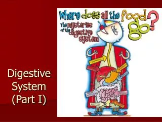
1 CHAPTER 23, part b DIGESTIVE SYSTEM. 2. Pharynx Food goes from mouth oropharynx esophagus Mucosa: inner wall is friction/abrasion resistant –stratified squamous epithelial tissue w/ mucous producing glands

E N D
1 CHAPTER 23, part b DIGESTIVE SYSTEM
2. Pharynx Food goes from mouth oropharynx esophagus Mucosa: inner wall is friction/abrasion resistant –stratified squamous epithelial tissue w/ mucous producing glands Muscularis Externica: 2 layers of skeletal muscles 1. longitudinal & 2. circular which propel food bolus into esophagus.
Esophagus • 10 inches long carries food from pharynx to stomach • Epiglottis closes of trachea when swallowing • Passes through mediastinum and pierces diaphragm, entering abdominal cavity, at the esophageal hiatus • Joins the stomach at cardiac orifice (opening) through cardiac sphincter
4. Esophagus Canal layers Mucosa – still non-keratinized stratified squamous epithelium Submucosa – mucus secreting glands to “grease” the food Muscularis externica: superior third skeletal; middle third mixed skeletal & smooth; inferior third all smooth Deglutition: Swallowing Serosa – Connective tissue?
6. Figure: 23.13; page 867 Cardiac Sphincter
8.( Inferior to?) stomach – temporary “storage tank” where chemical digestion of proteins start Chyme: food, when turned into a creamy paste Found in upper left quadrant 6-10 inches long Stretches! 50 ml to 4 liters (4000 ml) Rugae (wrinkly): longitudinal folds when empty
9. Regions of stomach Cardiac region (near heart) Fundus: dome shaped superior edge up against the diaphragm Body: Mid portion Pyloric region: distal end of stomach w/pyloric sphincter controlling entry to small intestine. Lesser curvature/greater curvature: inner, superior, curved edge/outer, inferior, curved edge
11. Omenta (pl): Double mesentery linings that help tether (hold) the stomach & other organs together and to the abdominal wall Lesser omentum: goes from liver to The stomach’s lesser curvature & then becomes continuous with stomach’s visceral peritoneum Greater omentum: stomach’s visceral peritoneum drapes off inferiorly, the Greater curvature where it continues to envelop the small intestine, the spleen and parts of the large intestine and finally attaches to the posterior abdominal wall
12. Microscopic Anatomy – four layers Epithelial (inner) - simple columnar Secretes: Protective alkaline mucus against stomach acids Gastric glands in deep gastric pits secrete gastric juices Parietal cells: HCl – hydrochloric acid Chief cells: Pepsinogen that becomes . Pepsin that starts protein digestion Enteroendocrine glands (gut hormones) chemical messengers to stimulate digestion activities
Muscularis: three layers instead of two • Longitudinal, circular and oblique • Mixes, moves and churns the food • Breaks it down into smaller pieces (more surface area) for chemical digestion • Bends into a “V” shape to force food into small intestine.
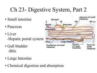
15. Small intestines – Main digestive organ Most digestion occurs here and all absorption occurs here About 20/7-13 feet long 1 to 1.6 inches in diameter 3 parts: duodenum, jejunum & ilium- all coiled up and “hanging” in mesentery
16. Duodenum – 10 inches long, coming off stomach at the pyloric sphincter. Bile duct & main pancreatic duct dump off bile and pancreatic juices in duodenum at the hepatopancreatic ampula and sphincter
18. Jejunum: Middle 8 feet Ileum: Final 12 feet that joins the large intestine at the ileocecal valve. Modifications for absorption – circular folds, villi and micro villi – increases surface area/absorption area 600 times – two tennis courts
19 Circular folds: longitudinal about 1 cm tall – set up turbulence that slows the chyme down for more absorption Villi: fingerlike projections – 1 mm high – velvety appearance – primarily absorptive, columnar epithelial tissue Dense capilary beds and lymphatic lacteals to absorb nutrients into blood and lymph
20 Microvilli – little fingerlike projections with even littler fingerlike projections. Fuzzy appearance so called a brush poarder
24. Endothelial: Simple columnar for absorption Goblet cells to secret mucous dissolves digesting molecules, keeps chyme liquid, allows for slippery surface Antimicrobial defenses Endothelial cells are sloughed off rapidly but, they reproduce just as quick
LIVER AND GALLBLADDER • Many metabolic and regulatory roles • In digestion it’s role is to produce bile • Bile is a fat emulsifier – breaks fats into tiny particles so its digested easier • Gallbladder stores the bile • Liver is largest gland in body – weighs about 3 lbs • Wedged shaped, upper right quadrant just under the diaphragm
Liver • Anatomy • Two main lobes, right and left, separated by falciform ligament • Also a quadrate and caudate lobe • Blood supply leaving the GI tract goes to the liver by the hepatic portal vein. This allows the liver to remove the “bad stuff” from the bad blood before it goes to the rest of the body
28. Histology Liver lobes are made up of liver lobules Six-sided, functional unit the size of a . sesame seed Each lobule is made up of plates of Hepatocytes (liver cells) which produce Bile and dumped into canaliculi which dump into bile ducts wich merge to form the common bile duct that joins with the cystic duct from the gallbladder to form the common bile duct that joins pancreatic duct hepatopancreatic ampula
31. Gallbladder Pear shaped sack on the posterior side of the liver near right lobe About 4 inches long Stores and concentrates bile. Ejects it into cystic duct to common bile duct into small intestine
32. Pancreas Secretes enzymes that are crucial to digestion It drains into the pancreatic duct that joins w/ the bile duct at the hepatopancreatic duct. “Tadpole” shaped, positioned laterally, mostly posterior of stomach, with large “head” side to right
34. Large Intestine About 5 feet long and almost 3 inches in diameter Main function is to absorb excess water from chyme/digested food and temporarily store the resedue and then eliminate them from the body as feces Joins with small intestine in lower right quadrant at the iliocecal valve
35. Ascending colon, cecum, appendix, splenic flexure, transverse colon, hepatic flexure, descending colon, sigmoid colon and rectum. Ending at anus with internal (involuntary) and external (voluntary) sphincter muscles Muscularis tunic (layer): 3 longitudinal rows of smooth muscle that pulls the intestinen making it “pucker” up into pouch-like segments called haustra (to draw up)
Mucosa tunic/layer • Simple columnar tissue. No absorption so, no folds or villi. Lots of crypts w/ goblet cells producing mucus to reduce friction on exiting feces.
Let’s go back to the liver A. Blood with oxygen comes from hepatic artery via the celiac trunk B. Blood with nutrients comes from hepatic portal vein • Bile is an emulsifier to digest fats for digestion and absorption. • Liver performs many other functions: • glucose to glycogen turn some amino acids into blood proteins turns ammonia into urea (urine) Kuppfer cells remove dead RBCs, bacteria, debris