Download
1 / 50
500 likes | 597 Views
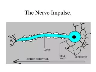
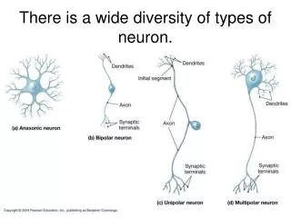
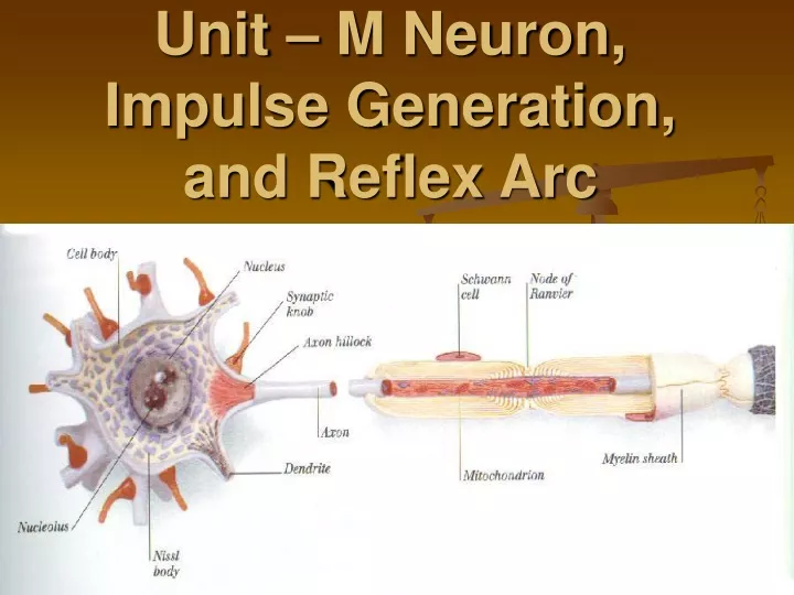
Unit – M Neuron, Impulse Generation, and Reflex Arc. Structures and Functions 1. Dendrites - Conduct a nerve impulse (message) towards a cell body. -Many dendrites enter a cell body. 2. Cell Body -Contains the nucleus and cell organelles

E N D
Structures and Functions 1. Dendrites -Conduct a nerve impulse (message) towards a cell body. -Many dendrites enter a cell body. 2. Cell Body -Contains the nucleus and cell organelles needed to keep the cell alive. -Relays impulse from Dendrite to Axon. 3. Axons -Conduct a nerve impulse away from the cell body.
4. Myelin Sheath -Protective coating of Schwann Cells around larger Axons 5. Nodes of Ranvier -Interrupted areas on the Myelin Sheath -Speeds up transmission of impulse. 6. Synaptic Terminal -Junction through which neurons signal to each other and to non-neuronal cells such as those in muscles.
Types of Neurons 1. Sensory Neuron (see animation) -Afferent Neuron: Moving away from a central organ or point. -Relays messages from receptors to the brain or spinal cord
2. Motor Neuron -Efferent Neuron: Moving toward a central organ or point. -Relays messages from the brain or spinal cord to the muscles and organs.
3. Interneuron (associated neuron or Connector Neuron) -Relays message from sensory neuron to motor neuron. -Make up the brain and spinal cord.
***A nerve is composed of long fibers of a number of Neurons***
Reflex Arc Reflexes are automatic, involuntary responses to changes occurring inside or outside the body. Some involve the brain (such as blinking the eye), while others do not (such as moving your hand away from a hot object). Why does the brain not have to be involved? If it were, by the time the impulse traveled to the brain, the brain figured out what was happening, and sent a response to the body, serious damage might occur.
Stages of Reflex Arc 1. Receptor is stimulated and formulate message. ie. nerve impulse 2. Sensory neuron takes the message to the Central Nervous System. (spinal cord) 3. Interneuron passes the message to a motor neuron.
4. Motor Neuron takes the message away from the C.N.S. to the effector. (muscle/organ) 5. The muscle receives the message and contracts. ***The brain finds out later what had happened***
Impulse Generation (Action Potential) Nerve impulses are electrical messages. If we measure the voltage of a resting neuron using a voltmeter, we will see a reading of –60 millivolts. Voltage is a comparison of electrical charge between two points.
When the neuron is stimulated, the charge changes briefly to +40 milivolts (mv), then back to –60mv. (-60mv means that the inside is 60mv more negative than outside). If we hook up our voltmeter to a machine called an oscilloscope, we can see the change in voltage over a period of time.
There is a difference in ion distribution on either side of the membrane of a neuron. At Rest -Na+ outside the Neuron -K+ and large negatively charged organic molecules inside the neuron.
The concentration of sodium ions Na+ is greater outside the axon than inside. • The concentration of potassium ions K+ is greater inside the axon than outside. • This unequal distribution is due to the sodium-potassium pump which actively transports Na+ out and K+ into the axon
The membrane is more permeable to K+ ions, and some K+ diffuses back while Na+ does not. • This unequal charge distribution, along with of the large negative molecules, causes the inside to be more negative than the outside. • This situation is called Resting Potential. -60mv
When the axon or dendrite is stimulated, sodium gates open which allows some Na+ to enter the axoplasm (interior). Now, the inside becomes more positive than the outside by 40 mv. This is called the Upswing Phaseof the action potential. The charge changes from –60 mv to +40 mv. The change is called Depolarization.
After the sodium gates have opened, then potassium gates open. K+exit the axoplasm. This is called the Downswing Phaseof the action potential. The charge returns to –60mv. The change is called Repolarization. **Note: Charge is back to normal, but ions are reversed
Finally, there is a Recovery Phase in which the sodium/potassium pump (ACTIVE TRANSPORT) returns Na+to the outside and K+to the inside. This is called the refractory period. During the refractory period, another action potential cannot be created.
After the recovery phase, the neuron is again at resting potential and can fire again!
Transmission of a signal – THINKING!!! • How is a signal transmitted down/along neuron? Think Dominoes!
Transmission of a nerve signal • Neuron has similar system • units are set up • once 1st is tripped, the rest fall in succession • all or nothing response • same force travels along neuron • have to re-set neuron to react again
So far we have only been looking at one point on the axon or dendrite. The opening of the sodium gates in one area causes the sodium gates in the next area to open because the sodium channels are voltage gated(sensitive to changes in voltage). We get a wave motion (chain reaction) moving down the nerve fiber.
1. RESTING POTENTIAL Charge is –60mv, -Na+ outside, K+ inside 2. STIMULUS REACHES THRESHOLD POTENTIAL “all or none response” 3. UPSWING OF ACTION POTENTIAL Depolarization, Na+ moves inside (sodium gates opened), charge from –60mv back to +40mv
4. DOWNSWING OF ACTION POTENTIAL Repolarization, K+ moves outside (Potassium gates opened), charge from +40mv back to –60mv 5. RECOVERY PHASE Sodium/Potassium Pump, moves Na+ out and K+ inside, charge is –60mv ****NOW NEURON CAN BE RE-STIMULATED **** **REMEMBER THIS IS A WAVE MOTION DOWN THE NEURON**
Myelin Sheath • Made from Schwann cells • insulate neuron • Increase speed of impulse • signal hops from node to node • “saltatory conduction” • 150m/sec (530km/hr)(vs. 5m/sec) unmyelinated signal direction myelinsheath
Multiple Sclerosis • T cells attack myelin sheath • loss of signal
Synapse (see video) Each axon branches off and ends with a swelled tip or terminal knob that lies close to but not touching the dendrite of another neuron. (or an organ). The entire region is called a synapse.
Transmission of nerve impulses across a Synaptic cleft is carried out by chemicals called Neurotransmitters. These substances are stored in vesicles at the end of the axon. Noradrenalin (speeds up activity) and acetylcholine (slows down activity) are examples of neurotransmitters.
When an impulse reaches the end of the axon like it usually would, not only does Na+ come into the axon, but Ca+2 as well since voltage gated calcium channels are opened. This calcium binds with contractile proteins that pull the neurotransmitter vesicles to the membrane surface. The vesicles join with the cell membrane, forcing the neurotransmitter into the cleft (exocytosis)
Neurotransmitter’s job is to increase the permeability of the sodium ions on the postsynaptic membrane. The Neurotransmitter binds to specific receptor sites on the dendrite of the next neuron. If enough transmitter substance is received, the neuron will “fire” and continue the impulse.
A neurotransmitter only has a short period to work once it has been released into the synaptic cleft. Enzymes rapidly break down the transmitter substance to clear the synapse so the next impulse can be transmitted. Monoamine oxidasebreaks down noradrenaline and Acetylcholinesterase breaks down acetylcholine.(see video)
Pain killers such as Tylenol act as an enzyme to break down the neurotransmitter to decrease the pain impulse. A natural painkiller in the body is prostaglandin. An impulse can only travel across a synapse in one direction. Only the axon contains neurotransmitter vesicles, so the impulse can only travel one way
AXON DENDRITE across a synapse. **** ALL OR NONE LAW (threshold): If enough neurotransmitter is received by the postsynaptic fiber, it will fire 100% (all). If not enough substance is received, it will not fire at all (none).
There are excitatory and inhibitory neurotransmitters in the body. When two excitatory neurotransmitters work together to cause an action potential, it is called summation.