Download
1 / 51
510 likes | 776 Views
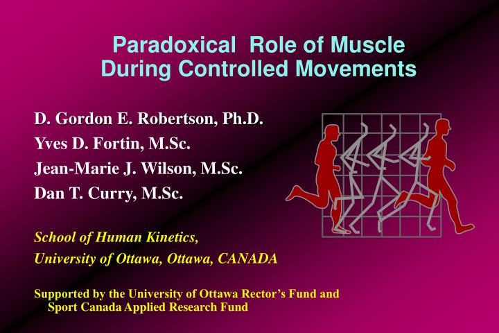
Paradoxical Role of Muscle During Controlled Movements. D. Gordon E. Robertson, Ph.D. Yves D. Fortin, M.Sc. Jean-Marie J. Wilson, M.Sc. Dan T. Curry, M.Sc. School of Human Kinetics, University of Ottawa, Ottawa, CANADA

E N D
Paradoxical Role of Muscle During Controlled Movements D. Gordon E. Robertson, Ph.D. Yves D. Fortin, M.Sc. Jean-Marie J. Wilson, M.Sc. Dan T. Curry, M.Sc. School of Human Kinetics, University of Ottawa, Ottawa, CANADA Supported by the University of Ottawa Rector’s Fund and Sport Canada Applied Research Fund
Introduction Anatomically muscle function is determined by examining how a muscle acts at a single joint during unloaded movements. For a biarticular muscle one joint is fixed while its function is determined at the other joint. The process is then repeated by reversing the fixation.
Introduction Cont’d Unfortunately, this method cannot consider how muscles function when several joints are moving simultaneously and how function is influenced by external loads and constraints. One scheme classifies muscle as either agonist, antagonist or stabilizer.
Paradoxical Role of Muscle Molbech suggested that biarticular muscle can act paradoxically during “controlled” or constrained movements. That is, when a limb is constrained at one or both ends, a flexor can produce extension or an extensor can produce flexion. This differs from Lombard’s paradox which considers doubly biarticular muscles at hip and knee during extension. Extension is explained by differential moment arms at knee and hip.
AX = AY Y K A X Molbech’s Model • A and K are hinges
AX = AY = AK + KH Y H K A X Molbech’s Model • A and K are hinges • H is constrained to Y-axis
AX = AY = AK + KH Y H O K A X Molbech’s Model • A and K are hinges • H is constrained to Y-axis • O is a muscle origin
AP = AK + KH Y P H O K A X Molbech’s Model • A and K are hinges • H is constrained to Y-axis • O is a muscle origin • extend line AK to P on arc of length AK+KH (limb length)
Molbech’s Model AP = AK + KH • A and K are hinges • H is constrained to Y-axis • O is a muscle origin • extend line AK to P on arc of length AK+KH (limb length) • draw ray L from P through O Y P H O K L A X
AP = AK + KH Y P H O K G L A X Molbech’s Model • A and K are hinges • H is constrained to Y-axis • O is a muscle origin • extend line AK to P on arc of length AK+KH (limb length) • draw ray L from P through O • if ray OG is above L muscle causes extension
AP = AK + KH Y P H O K G L A X Molbech’s Model • A and K are hinges • H is constrained to Y-axis • O is a muscle origin • extend line AK to P on arc of length AK+KH (limb length) • draw ray L from P through O • if ray OG is below L muscle causes flexion
Molbech’s Model AP = AK + KH • A and K are hinges • H is constrained to Y-axis • O is a muscle origin • extend line AK to P on arc of length AK+KH (limb length) • draw ray L from P through O • if ray OG is along L muscle acts isometrically Y P H O G K L A X
Paradoxical Muscle Activity? • what factors characterize paradoxical activity? • how are muscle contributions determined? • no means of directly measuring multiple muscle forces, simultaneously and in vivo
Classification of Muscle Functions Muscle functions as: agonist, antagonist, stabilizer, extraneous or paradoxical. Function is characterized by: • its anatomical function (flexor, extensor, . . .) • the joint’s kinematics (flexion, extension, . . .) • the joint’s kinetics (moment of force) • its activity level (EMG above a threshold) • its kinematics (lengthening or concentric, shortening or eccentric, static or isometric)
Rectus Femoris during Hip Flexionin Soccer Kicking • hip flexor and knee extensor • hip flexing • hip flexor moment • active contraction • concentric, shortening Classified: concentric agonist
Gluteals during Hip Extensionin Hurdling • hip extensor • hip extending • extensor moment • active contraction • concentric, shortening Classified: concentric agonist
Erector Spinae while Stooped at Waist • trunk extensor • trunk statically extended • extensor moment • active co/contraction • isometric Classified: isometric agonist
Assumptions It was assumed that, in skilled athletes, muscles were only recruited if they were beneficial to the movement. Extraneous or inappropriate contractions and cocontractions were assumed to be nonexistent.
Classification of Muscle Function as Paradoxical To determine paradoxical activity the following must occur: • joint motion must be opposite to muscle’s anatomical function • net moment of force at the joint must be opposite to anatomical function • muscle must be active • muscle must be shortening
Methods - Joint Kinematics The joint motion must be opposite to the accepted anatomical role of the muscle. For example, for the biarticular hip flexor, rectus femoris, to act paradoxically at the hip then the hip must be extending.
Methods - Joint Kinetics The moment of force at a joint must be opposite to the usual anatomical role of the muscle. For example, the moment of force at the knee must be extensor for a knee flexor, such as, a hamstring or gastrocnemius muscle, to be considered paradoxical.
Methods - Muscle Activity (EMG) The muscle must be actively contracting. • criterion for significant muscle activity was: muscle EMG level was >25% EMGMVC (i.e., during a maximal voluntary contraction, MVC) • EMG level of less than 25% EMGMVC was considered too low for the muscle to contribute significantly to the movement
Methods - Muscle Kinematics The muscle must be contracting concentrically, that is, it must be shortening. An isometric or an eccentric contraction of the muscle would imply a stabilizing or antagonist function.
Purpose - Squat Lift The purpose of this study was to determine the functional role of eight leg muscles, four of which are biarticular, during knee extension of loaded and unloaded squat lifting.
Squat Lift • six experienced male weight-lifters • cinefilm at 50 frames/second • Kistler force platform • 80% maximum lift and no load conditions • EMGs from muscles: rectus femoris, gluteus maximus, biceps femoris, semitendinosus, vastus lateralis, gastrocnemius, tibialis anterior and soleus • muscle lengths from Frigo’s and Pedotti’s model with modifications by Hubley
Conclusions - Squat Lift • biarticular muscles acted as stabilizers of knee • no paradoxical activity was performed • prime movers were the monoarticular muscles • knee muscles were responsible for starting the descent and preventing hyperextension at the end of the lift
Purpose - Ergometer Rowing • The purpose of this study was to determine the functional role of six leg muscles, three of which are biarticular, during drive phase of ergometer rowing.
Experimental Set-up Strain gauge force transducer Gjessing ergometer Flywheel Workload adjustment Foot stretcher Biological amplifier Kistler force platform Charge amplifier Bridge amplifier A/D converter Cinecamera Microcomputer
Methods • four female and four male elite rowers performed on the ergometer while kinematic information was recorded on cinefilm at 50 frames/second • workload 25 N (2.6 kp) for females, 30 N (3.1 kp) for males • ten cycles at 27 strokes/minute • EMGs from MVCs recorded separately
Methods - Forces • forces measured by Kistler force platform angled at 10 degrees • sampled at 200 Hz • rotated to Newtonian frame of reference • handle forces measured by in-line force transducer
Methods - Joint Kinetics • inverse dynamics to compute the net moments of force produced at the ankle, knee and hip joints • powers produced by moments were computed as product of moment and joint angular velocity (M . w)
Methods - Electromyography • muscles investigated were the monoarticular: vastus lateralis, soleus, gluteus maximus and the biarticular: biceps femoris, rectus femoris and gastrocnemius • sampled simultaneously at 200 Hz for ten rowing cycles • CMRR >100 dB, 10-700 Hz filter, 2000 gain • linear envelope detector (6 Hz cutoff) • ensemble averaged across entire stroke
Methods - Muscle Length • instantaneous length of each muscle was computed from the digitized image with to adaptation of the muscle length model of Frigo and Pedotti (1978) • model based on marker kinematics and subject height
Results To provide higher accuracy, the drive phase was divided in three equal parts; each criterion delineated by the classification scheme was verified for each muscle during each third of the drive.
Moment Powers of Male Rowers 1500 Hip RMBS RMDH 1000 RMKH RMNT RMSD 500 Concentric 0 -500 Eccentric Knee 1000 Power (watts) 500 Concentric 0 -500 Eccentric Ankle 500 Concentric 0 Eccentric -500 0 10 20 30 40 50 60 70 80 90 100 Time (% drive)
1500 Hip RFEK RFLM 1000 RFLN RFMM 500 Concentric 0 -500 Eccentric Knee 1000 Power (watts) 500 Concentric 0 -500 Eccentric Ankle 500 Concentric 0 Eccentric -500 0 10 20 30 40 50 60 70 80 90 100 Moment Powers of Female Rowers Time (% drive)
Results - Knee • angular velocity curve portrays relatively slow joint motion (below 4 rad/s) • knee moment was extensor for 2/4 females and all males • weak moment for females (>100 N.m) but strong for males (>150 N.m) • moment produced positive work for all males during middle and late drive and for two females during middle drive
Results - Knee Kinetics Figure is from a male subject and depicts the knee’s angular velocity, net joint moment of force and the power produced by the moment of force.
Results - EMG • EMG curves show that most muscles contracted in-phase with each other • most were significantly active (EMGmax >25% EMGMVC) during middle and late third of drive • most were below threshold during first third of drive (i.e., end of recovery)
Results - Muscle Shortening • muscle velocities were time normalized to the drive duration • positive velocity indicated shortening or concentric contraction (same convention as muscle physiologists) • negative velocity indicated lengthening or eccentric contraction
Discussion - Classification • biceps femoris acted paradoxically for knee extension in one female and two males • gastrocnemius acted paradoxically for knee extension in one female and two males • rectus femoris acted paradoxically for hip extension in three of four males and three of four females during late drive
Discussion - Powers • female rowers produced majority of work by hip extensors; knee and ankle extensors produced insignificant amounts of work or negative work • male rowers had contributions from ankle, knee and hip extensors; mainly from hip
Conclusions - Rowing Paradoxical activity was detected infrequently during knee extension by the biarticular biceps femoris and gastrocnemius. More intriguing was the detection of paradoxical activity from the action of rectus femoris during hip extension.
References Carlsoo, S. 1966 Acta Morphologica Neerlando-Scandinavica, 6:377-386 Elftman, H. 1939 American Journal of Physiology, 125:357-366 Frigo, C & Pedotti, A. 1978 Biomechanics IV, 355-360 Lombard, WP. 1903 American Physical Education Review, 8:141-145 Molbech, S. 1965 Acta Morphologica Neerlando-Scandinavica, 6:171-178 Rasch, PJ & Burke, RK. 1978 Kinesiology and Applied Anatomy, 6th edition
Questions? Answers? Thank you.
Pectoralis major during horizontal flexion in tennis forehand stroke • horizontal flexor • horizontal flexing • horizontal flexor • active contraction • shortening Classified: concentric agonist
Anterior deltoids during arm stand in gymnastics • shoulder flexor • shoulder statically extended • extensor moment • active cocontraction • isometric Classified: stabilizer
Hamstrings during knee extension in running • knee flexor • knee extending • flexor moment • active contraction • eccentric, lengthening Classified: eccentric agonist
Neck extensors during keyboarding • neck extensor • neck statically flexed • extensor moment • active contraction • isometric Classified: isometric agonist
Rectus abdominis during trunk flexion in high jumping • trunk flexor • trunk flexing • flexor moment • active contraction • concentric, shortening Classified: concentric agonist