Download
1 / 59
600 likes | 677 Views
Radiation therapy. Radiation therapy Radiotherapy Radiation oncology.

E N D
Radiation therapy
Radiation therapy Radiotherapy Radiation oncology
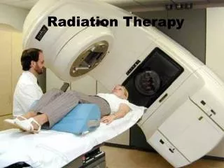
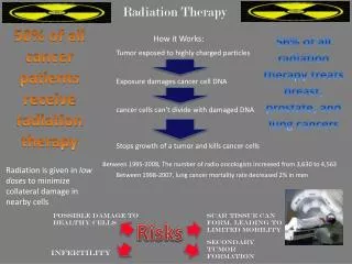
Radiation therapy (also radiotherapy or radiation oncology, sometimes abbreviated to XRT) is the medical use of ionizing radiation as part of cancertreatment to control malignantcells (not to be confused with radiology, the use of radiation in medical imaging and diagnosis). Radiotherapy may be used for curative or adjuvant cancer treatment. It is used as palliative treatment (where cure is not possible and the aim is for local disease control or symptomatic relief) or as therapeutic treatment (where the therapy has survival benefit and it can be curative).
Total body irradiation (TBI) is a radiotherapy technique used to prepare the body to receive a bone marrow transplant. Radiotherapy has several applications in non-malignant conditions, such as the treatment of trigeminal neuralgia, severe thyroid eye disease, pterygium, pigmented villonodularsynovitis, prevention of keloid scar growth, and prevention of heterotopic ossification. The use of radiotherapy in non-malignant conditions is limited partly by worries about the risk of radiation-induced cancers.
Radiotherapy is used for the treatment of malignant tumors (cancer), and may be used as the primary therapy. It is also common to combine radiotherapy with surgery, chemotherapy, hormone therapy or some mixture of the three. Most common cancer types can be treated with radiotherapy in some way. The precise treatment intent (curative, adjuvant, neoadjuvant, therapeutic, or palliative) will depend on the tumour type, location, and stage, as well as the general health of the patient.
The therapeutic use of IR is based on it’s biological action, that is, it’s ability to cause changes in cells, tissues, organs, the body as a whole. It depends on the ABSORBED DOSE (AD) – e n e r g y transmitted to irradiated tissues (Gy). Clinical-Biological Bases of RT of Tumors
To spare normal tissues (such as skin or organs which radiation must pass through in order to treat the tumour), shaped radiation beams are aimed from several angles of exposure to intersect at the tumour, providing a much larger absorbed dose there than in the surrounding, healthy tissue.
Dose The amount of radiation used in radiation therapy is measured in gray (Gy), and varies depending on the type and stage of cancer being treated. For curative cases, the typical dose for a solid epithelial tumor ranges from 60 to 80 Gy, while lymphoma tumors are treated with 20 to 40 Gy.
Fractionation The total dose is fractionated (spread out over time) for several important reasons. Fractionation allows normal cells time to recover, while tumor cells are generally less efficient in repair between fractions. Fractionation also allows tumor cells that were in a relatively radio-resistant phase of the cell cycle during one treatment to cycle into a sensitive phase of the cycle before the next fraction is given. Similarly, tumor cells that were chronically or acutely hypoxic (and therefore more radioresistant) may reoxygenate between fractions, improving the tumor cell kill.
Types of radiation therapy Historically, the three main divisions of radiotherapy are external beam radiotherapy (EBRT or XBRT) or teletherapy, brachytherapy or sealed source radiotherapy, and systemic radioisotope therapy or unsealed source radiotherapy. The differences relate to the position of the radiation source; external is outside the body, brachytherapy uses sealed radioactive sources placed precisely in the area under treatment, and systemic radioisotopes are given by infusion or oral ingestion. Brachytherapy can use temporary or permanent placement of radioactive sources.
The temporary sources are usually placed by a technique called afterloading. In afterloading a hollow tube or applicator is placed surgically in the organ to be treated, and the sources are loaded into the applicator after the applicator is implanted. This minimizes radiation exposure to health care personnel. Particle therapy is a special case of external beam radiotherapy where the particles are protons or heavier ions. Introperative radiotherapy is a special type of radiotherapy that is delivered immediately after surgical removal of the cancer. This method has been employed in breast cancer (TARGeted Introperative radioTherapy), brain tumours and rectal cancers.
Particle Therapy In particle therapy (Proton therapy), energetic ionizing particles (protons or carbon ions) are directed at the target tumor.The dose increases while the particle penetrates the tissue, up to a maximum (the Bragg peak) that occurs near the end of the particle's range, and it then drops to (almost) zero. The advantage of this energy deposition profile is that less energy is deposited into the healthy tissue surrounding the target tissue.
Principles of Radiotherapy 1. Delivering of an optimal dose to the tumor 2. Minimal damage of surrounding organs & tissues. 3. Measures stimulating protective forces of the body
Radioisotope Therapy (RIT) Systemic radioisotope therapy is a form of targeted therapy. Targeting can be due to the chemical properties of the isotope such as radioiodine which is specifically absorbed by the thyroid gland a thousand fold better than other bodily organs. Targeting can also be achieved by attaching the radioisotope to another molecule or antibody to guide it to the target tissue. The radioisotopes are delivered through infusion (into the bloodstream) or ingestion. Examples are the infusion of metaiodobenzylguanidine (MIBG) to treat neuroblastoma, of oral iodine-131 to treat thyroid cancer or thyrotoxicosis, and of hormone-bound lutetium-177 and yttrium-90 to treat neuroendocrine tumors (peptide receptor radionuclide therapy). Another example is the injection of radioactive glass or resin microspheres into the hepatic artery to radioembolize liver tumors or liver metastases.
Today more than 70 % of oncological patients obtain radiation treatment • as initial • in connection with relapses of disease • with the palliative purpose
Treatment of tumors may be: RADICAL RT– complete cure of the patients –Indications: an early stages (I-II). PALLIATIVE RT - a temporary improvement of patients life, life prolongation (stages III- IV). SYMPTOMATIC RT - to alleviate the most severe manifestations of the disease
Approximately 45 % are cured 22 – surgery (independently or in combination) 18 – radiation therapy (independentlyor as a leading method) 5 – chemotherapy (independentlyor, more often, it is combined with surgery and radiation therapy)
The role of a radiotherapy grows because of early diagnostics of oncological diseases
Radiotherapeutic Interval Selection of an irradiation regimen: delivery of an optimal total tumor dose (TTD) Radiosensitivity of healthy and tumor cells is practically the same. The radiotherapeutic interval is a difference between radiosensitivity of the tumor and surrounding healthy organs and tissues It should be increased
Mechanisms of cells’ death • interphase death (apoptosis) • loss of reproductive ability
Factors of modification • Natural radiosensitivity • Oxygen • Cellular restitution • Restitution of potentially lethal damages • Cellular cycle • Cellular proliferation
Methods of radiomodification Oxygenous effect: Oxybaroradiotherapy Hypoxiradiotherapy- gaseous hypoxia-protection of normal tissues Chemical compounds: Radiosensibilizers Hyperthermia Polyradiomodification
Organs of hazard Normal tissues, which radiosensitiveness can essentially influence on planning of treatment and/or the appointed dose
Effect of a radiotherapy depends on a dose in a tumour
Factors of therapeutic dose limitation • tolerance of normal tissues around a tumour, • that is • acute responses of tissues • late radiation effects
Radiations for radiotherapy • photons • electrons • beta particles • heavy particles • (protons, neutrons)
Sources of radiotherapeutic radiations • X-Rays • linacs • cyclic accelerators • nuclear reactors • radionuclides
On distance Source – Target • Distant irradiation • Contact irradiation • (Brachytherapy)
On localization of source • outside irradiation • intracavitary irradiation • interstitial irradiation • enteral or parenteral introducing of radionuclides
On type of apparatus • Roentgenotherapy (X-Rays) • (low voltage, orthovoltage) • Telegammatherapy • Therapy by photons of a linac • Therapy by electrons of a linac • Brachytherapy by the closed radionuclides • Brachytherapy by "grains" of radionuclides
Methods of patient’s irradiation S t a t i c irradiation can be performed through: Single field technique Multiple field technique M o b i l e irradiation: Rotational
Contraindications for RT grave patient’s state, cachexia, anemia, leukopenia, acute septic states decompensated states of heart, liver, kidneys Active tuberculosis Extension of tumors to adjacent hollow organs, growth into great blood vessels. An inflammatory process
RT of NON-TUMOR DISEASES It is carried out only according to strict indications when other methods failed Single and total doses should be small in the range of 0.1-0.7 Gy every alternate day up to a TFD of 4-7 Gy The basic method is a direct local irradiation of the focus using the remote technique Radiation field is equal to the focal size The X-ray unit or telegammatherapy is used at deep foci (more often X-ray therapy )
Inflammatory processes We cause local plethora, an increased permeability of capillaries, formation of bioactive substances, lymphocytolysis The principle of treatment – the severe the process, the lower the dose. At the initial stage, it is possible to terminate the process, alleviate pain, relieve inflammation. At phase of necrosis and purulation - rapid infiltrate melting and its delimitation. At the phase of regeneration – rapid wound epithelisation.
Mechanisms of activity • Anti-inflammatory • Anesthesia • Desensitization • Ablation of function
Purulent surgical infection • Purulent wounds • Pain pancreatitis • Postoperative anastomosites • Thrombophlebites • Arthritises and arthroses • Bursitises • Neuritises • Neuralgias • Ablation of a hyperfunction
Clinical-dosimetric planning of RT The main clinical-dosimetric task is to create the most favorable spatial distribution of absorbed radiation doses in patient’s body EXTERNAL BEAM THERAPY