Download
1 / 18
300 likes | 1.67k Views
BASIC 12 LEAD ECG INTERPRETATION. Dr. Jeffrey Elliot Field, HBSc. DDS, Fellow, American Dental Society of Anesthesia Diploma, the National Dental Board of Anesthesia. 1. 8/7/2014. Objectives . 2. To gain a cursory understanding of 12 lead ECG ’ s. 8/7/2014.

E N D
BASIC 12 LEAD ECG INTERPRETATION Dr. Jeffrey Elliot Field, HBSc. DDS, Fellow, American Dental Society of Anesthesia Diploma, the National Dental Board of Anesthesia. • 1 8/7/2014
Objectives • 2 To gain a cursory understanding of 12 lead ECG’s 8/7/2014
12 lead ECG’s give you the opportunity to look for all the arythmias we have studied in a 360 degree view of the heart with the added bonus of being able to diagnose and localize myocardial infarctions ( i.e. areas of muscle damage) • Remember to look at all leads to rule out all of the arrhythmias and abnormalities we have studies thus far. • In particular look first at Leads I , II, and III as these will be the most familiar to you .
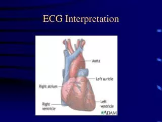
In a 12 lead ECG 10 electrodes/leads attached to the patient.
Myocardial Infarction • Myocardial infarctions can be categorized as follows: • -Q-wave MI • -Non Q-wave MI
Q-Wave Myocardial Infarction • This is the classic presentation for MI’s. • The developing MI is seen as ST segment elevation followed by deepening Q-waves in the leads where ST segment elevation was 1st seen. • The ECG changes are accompanied by elevated cardiac enzymes and markers and of course physical signs and symptoms of an MI ( chest pain ,nausea ,vomiting , etc)
Non Q-Wave Myocardial Infarction • In this case you get classic signs and symptoms symptoms of an MI(i.e. elevated cardiac enzymes and markers and of course physical signs of an MI ( chest pain ,nausea ,vomiting , etc) But non of the usual ECG changes ( i.e. ST segment elevation and deepening Q-waves). In fact sometimes the only clue on the ECG are inverted T-waves.
Cardiac Enzyme changes and Markers for MI • There are 4 markers for cardiac enzymes as follows: • CK-MB isoenzyme • CK-MB isoforms • Myoglobin • Troponin T or Troponin I
Relative Advantages and Disadvantages of the Various cardiac Markers
Therefore the best test overall is Troponin T or I. But these will not detect reinfarction and therefore more than one test is required. The current recommendation is to combine one of the CK-MB tests with one of the Troponin tests. It should be noted that high troponin levels post MI correlate with poor outcomes. Finally please note CK-MB also rises in unstable angina ( damaged cells that will recover ) as well as MI ( damaged cells that won’t recover) so it won’t differentiate between unstable angina and an MI
Localizing Myocardial infarctions • Anterior( blockage of left anterior descending artery) –look for ECG changes in leads V1-V4 • Inferior ( blockage of right coronary artery or less commonly right circumflex)- look for ECG changes in leads II, III, and AVF
Localizing Myocardial infarctions continued • Lateral Infarction ( blockage of circumflex or diagonal branch of the LAD)-look in leads V5, V6 and AVL • Posterior (blockage of right coronary artery or circumflex) –look for mirror image changes to anterior in V1-V4 (i.e. ST depression and dominant R-wave).