Download
1 / 67
670 likes | 905 Views
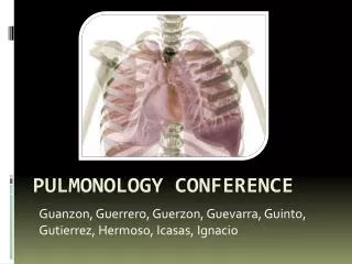
Guanzon , Guerrero, Guerzon , Guevarra , Guinto , Gutierrez. Pulmonology Conference. General Data. LS 1 month old / Male Lives in Malabon City Roman Catholic Single Informants: mother and grandmother Good reliability. Chief Complaint: Difficulty of Breathing.

E N D
Guanzon, Guerrero, Guerzon, Guevarra, Guinto, Gutierrez Pulmonology Conference
General Data • LS • 1 month old / Male • Lives in Malabon City • Roman Catholic • Single • Informants: mother and grandmother • Good reliability
Chief Complaint: Difficulty of Breathing
(+) productive cough (-) fever, colds, vomiting, diarrhea, anorexia No consult was done. No medication was given. 7 days PTA (+) productive cough (-) fever, colds Consult at a Pediatric clinic Medication: Ambroxol, 0.25ml TID taken for 2 days No relief of symptoms 6 days PTA 4 days PTA (+) difficulty of breathing, (+) fever (38.5 ⁰C) (+) productive cough (-) colds Consult at a public hospital Medication Salbutamolnebulization TID for 5days 0.65% NaClnasal solution with suctioning Cefixime(unrecalled dose; #/day?; for how long?) Compliant relief of difficulty of breathing & lysis of fever
(+) recurrence of difficulty of breathing (+) productive cough (-) fever Salbutamolnebulization(how many times?) Relief? 1 day PTA 10 hours PTA (+) recurrence of difficulty of breathing (+) productive cough (-) fever Consult at a pediatric clinic with an assessment of bronchial asthma and was advised admission. ADMISSION
Review of Systems • General: (-) weight change, (-) loss of appetite • Cutaneous: (-) rash • Heent: (-) excessive lacrimation, (-) epistaxis, (-)excessive salivation, • (-) nasal structures, • Cardiovascular: (-)cyanosis, (-) fainting spells • Respiratory: (-) cough, • Gastrointestinal: (-) nausea, vomiting, (-)constipation, (-)abdominal pain • Genito-urinary: (-) frequency,(-)hematuria • Nervous/Behaviour: (-) convulsions, stiffness • Musculoskeletal: (-) joint swelling, (-) limitation of motion, (-)limping • Hematopoietic: (-)pallor, (-) abnormal bleeding, (-) easy bruisability
Gestational History • The patient was born term to a 20 year old primigravid. Mother had regular prenatal checkups. She denied smoking, drinking of alcoholic beverages, and use of illegal drugs during the pregnancy. Mother calimed non-exposure to chemicals, radiation, and viral exanthems. No complications noted during pregnancy.
Birth History • The patient was born term via CS, singleton, with no complications during and after delivery.
Neonatal History • The patient had spontaneous respiration, no pallor, no cyanosis, no jaundice and good cry. There was no note of gross congenital anomaly. The primary caregiver is the mother.
Feeding History • The patient was breastfeeding for 3 days. From 4th day the patient started formula feeding,Bona (2 oz of water and 1 scoop) for one month and shifted to Nan (2-4 ounces/feeding) since Jan. 17, 2010. Patient is currently on Nan (2-4oz/feeding).
Developmental Milestones • Can raise head slightly • Hands fisted • Eyes follow objects midline • Has throaty, gurgling sound • Regards face • => at par with age
Past Medical History • No previous admission • (-) allergy, (-) DM, (-) HPN, (-) cardiac/ renal disease (-) chickenpox, (-) measles, (-) mumps, • No previous surgery and transfusion
Immunization History • (+) BCG 1 • (+) HepaB 1
Family History • (+) HPN – maternal grandmother • (+) allergies – father(to seafood) • (-) Asthma • (-) PTB, • (-) DM • (-) Cancer
Socioeconomic & Environmental History • The patient lives with her mother, maternal grandmother and aunts and uncles in the maternal grandmother’s house, cemented, well-lit, well ventilated. The patient’s room is not-airconditioned. Water for consumption was district water supply. Garbage was collected daily. Patient has no exposure to cigarette smoke. No pets in the house. No factories nearby.
Physical Examination • Awake, alert, in respiratory distress, well-nourished, well-hydrated • VS: HR 124 bpm regular, RR 55/min, T 36.5 C, Wt: 4.38 kg Z(0)Ht 58 cm Z( below +2), WFH: Z(-2) • Warm and moist skin, no active dermatosis • No head deformities, anterior and posterior fontanels not depressed or bulging • Pink palpebralconjunctivae, anictericsclerae, eyeballs not sunken • Non-hyperemic EAC, TM intact, AU • Nasal septum midline, (+) nasal discharge, turbinates not congested • Moist buccal mucosa, tonsils not enlarged, non-hyperemic PPW
Physical Examination • Supple neck, (-) palpable cervical lymph nodes • Symmetrical chest expansion, (+) subcostal retraction, (+) crackles on both lung fields • Adynamicprecordium, AB at 4th LICS MCL, (-) murmur, • Globular abdomen, NABS, no organomegaly, no mass • Full and equal pulses on all extremities, (-) cyanosis
Neurologic Examination • Awake, alert, responds to stimuli • Pupillary reflex 1-2mm ERTL, no facial asymmentry(+) corneal reflex • (-) Tremor, abnormal limb movement • Good muscle tone, bulk, no flaccidity rigidity, spasticity • (-) Meningeal signs
Salient Features • Pertinent Negatives • (-) FH of asthma • (-) Cyanosis • Pertinent Positives • Difficulty of breathing • 1-day high-grade fever • FH of atopy • Productive cough • Nasal discharge • Tachypnea • Subcostal retractions • Crackles
Fever, productive cough, retractions, crackles on both lung fields Differential Diagnosis
Infectious • Pneumonia • Upper Respiratory Tract Infection • Lower Respiratory Tract Infection • Non infectious • Bronchial Asthma
Bronchial asthma • Reversible obstructive lung disease • Cough, dyspnea, recurrent wheeze worsening at night • Mild, moderate or severe • Relieved by inhaled B2 agonist
URTI • Croup • Etiologic agent: viral (Parainfluenza) • Rhinorrhea, pharyngitis, mild cough, low grade fever for 1-3 days before signs & symptoms of upper airway obstruction becomes evident • Barking cough, hoarseness, inspiratorystridor, tachypnea, retractions • Improves with epinephrine & steroids
Tracheitis • Caused by Staphylococcus aureus&Haemophilus influenza • Gradual progression of respiratory distress over 2-3 days accompanied by high fever • Subglottic narrowing • Unresponsive to racemic epinephrine • Treated with antibiotics
LRTI • Acute bronchiolitis • Results from inflammatory obstruction of the small airways • Caused by RSV, parainfluenza, adenovirus • Mild URTI, decreased appetite, fever, paroxysmal wheezy cough, dyspnea, irritability • Tachypnea, nasal flaring, retractions, fine crackles, prolonged expiratory phase • Hyperinflated lungs with patchy atelectasis
Pulmonary Tuberculosis • Signs & Symptoms : • Cough of 2 weeks or more • Failure to return to normal health after an infection • Painless cervical lymphadenopathy • Weight loss or poor weight gain • Failure to respond to appropriate antibiotic therapy
Pneumonia • Common causes: H. influenzae, S. pneumoniae • Pneumonia • Fast breathing, no chest indrawing • Severe Pneumonia • Fast breathing, chest indrawing • No central cyanosis • Very Severe Pneumonia • Central cyanosis, inability to feed or drink, stridor, convulsions, lethargic, severe undernutrition
Impression on Admission • t/c Pneumonia
Pneumonia • Pneumonia is a severe form of acute lower respiratory infection that specifically affects the lungs. • Two tell-tale symptoms of pneumonia: • fast breathing • difficulty of breathing WHO & UNICEF: Pneumonia – the forgotten killer of children; 2006
Pneumonia • Characterized by inflammation of the alveoli and terminal airspaces in response to invasion by an infectious agent introduced into the lungs through hematogenous spread or inhalation. • The inflammatory cascade triggers the leakage of plasma and the loss of surfactant, resulting in air loss and consolidation. This is in contrast to pneumonitis, which is caused by noninfectious agents such as radiation or chemicals. Pneumonia: eMedicine 2009
Increased Risk for Pneumonia • Intubation, tracheostomy, impaired cough reflex, and aspiration: • provide infectious organisms with easier access to the alveoli and terminal airspaces. • Ciliarydyskinesia, bronchial obstruction, viral infection, cigarette smoke, and certain chemical agents: • These conditions create disruption in the mucociliary blanket. • Anatomic abnormalities (eg, sequestrations), gastric fluid aspiration or other causes of noninfectious inflammation, altered pulmonary blood flow, and pulmonary edema: • These conditions increase the predisposition for pneumonia. • Immunodeficiency and immunosuppression: • These conditions increase predisposition for pneumonia. Pneumonia: eMedicine 2009
Infections • Viral infections • Accumulation of mononuclear cells in the submucosa and perivascularspace partial obstruction of the airway • wheezing and crackles. • Alveolar type II cells lose their structural integrity and surfactant production is diminished, a hyaline membrane forms, and pulmonary edema develops • Fungal infections • Unusual and are typically found in patients with inadequate immune function • Diffuse infiltrate of organisms or focal areas of fungal growth. • Appear ill and may have more subtle physical findings Pneumonia: eMedicine 2009
Infections • Bacterial infections • The alveoli fill with proteinaceous fluid, which triggers a brisk influx of RBCs and polymorphonuclear cells (red hepatization) followed by the deposition of fibrin and the degradation of inflammatory cells (gray hepatization). • Resolution: intra-alveolar debris is ingested and removed by the alveolar macrophages consolidation decreased air entry and dullness to percussion • Inflammation in the small airways leads to crackles. Wheezing is less common than in viral infections. • The inflammation and pulmonary edema that result from these infections cause the lungs to become stiff and less distensible, thereby decreasing tidal volume. The patient must increase his or her respiratory rate to maintain adequate ventilation. • Poorly ventilated areas of the lung may remain well perfused, resulting in ventilation/perfusion (V/Q) mismatch and hypoxemia. Tachypnea and hypoxia are common. Pneumonia: eMedicine 2009
Canadian Medical Assoc J: A practical guide for the diagnosis and treatment of pediatric pneumonia; 1997
Academy of American Family Physicians: CAP in Infants & Children; 2004
Pneumonia Diagnosis • Chest X-rays and laboratory tests are used to confirm the presence of pneumonia, including the extent and location of the infection and its cause • Clinical symptoms • Children and infants are presumed to have pneumonia if they exhibit a cough and fast or difficult breathing. WHO & UNICEF: Pneumonia – the forgotten killer of children; 2006
Pneumonia Transmission • Aspiration • Pathogens already present in a child’s nose or throat and are then inhaled into the lungs, causing infection. • Contaminated air droplets • Blood-borne infections • Neonates - birth canal or from contaminated substances contacted during delivery WHO & UNICEF: Pneumonia – the forgotten killer of children; 2006
Risk Factors • Undernourished children • Not exclusively breastfed • Inadequate zinc intake, are at • higher risk of developing pneumonia • Other illnesses, AIDS or measles, • Living in crowded homes • Exposure to parental smoking or indoor air pollution WHO & UNICEF: Pneumonia – the forgotten killer of children; 2006
WHO & UNICEF: Pneumonia – the forgotten killer of children; 2006
In developing countries acute respiratory infections cause up to 5 million deaths annually among children less than 5 years old. • Several risk factors increase the incidence or severity of pneumonia in children: prematurity, malnutrition, low socioeconomic status, passive exposure to smoke and attendance at day-care centres.10 Underlying disease, especially that affecting the cardiopulmonary, immune or nervous systems, also increases the risk of severe pneumonia Canadian Medical Assoc J: A practical guide for the diagnosis and treatment of pediatric pneumonia; 1997
Mechanism of Symptoms • In order to identify very sick children with cough or difficult breathing one checks two clinical signs: fast breathing and chest indrawing. When children develop pneumonia, their lungs become stiff. One of the body’s responses to stiff lungs and hypoxia is fast breathing. When the pneumonia becomes more severe, the lungs become even stiffer. Chest indrawing may develop. Its presence is a sign of severe pneumonia.
Clinical Assessment • Pneumonia can be defined clinically as the presence of lower respiratory tract dysfunction in association with radiographic opacity. • WHO has promoted an algorithm to assess children who present with cough and fever. • Tachypnea, considers an increased respiratory rate • >50 breaths/min in infants • >40 breaths/min in children >11 months • Suprasternal, subcostal or intercostalretractions indicates greater severity. Canadian Medical Assoc J: A practical guide for the diagnosis and treatment of pediatric pneumonia; 1997
Academy of American Family Physicians: CAP in Infants & Children; 2004
Radiographic confirmation is considered the gold standard. • However, no finding in itself can be used to diagnose or rule out pneumonia. The absence of the symptom cluster of respiratory distress, tachypnea, crackles and decreased breath sounds accurately (100% specificity) excludes the presence of pneumonia (level II evidence). • Assessment of oxygenation gives a good indication of the severity of disease. • Oximetry should be considered in the assessment of a child with suspected pneumonia and in all children admitted to hospital with pneumonia, because the results correlate well with clinical outcome and length of hospital stay (level II evidence). Canadian Medical Assoc J: A practical guide for the diagnosis and treatment of pediatric pneumonia; 1997
Two classic presentations have been described for pneumonia: • Typical pneumonia: fever, chills, pleuritic chest pain and a productive cough. • Atypical pneumonia: gradual onset over several days to weeks, dominated by symptoms of headache and malaise, nonproductive cough and low-grade fever. • Unfortunately, the overlap of microbial agents responsible for these presentations thwarts identification of the causal pathogen on the basis of clinical presentation. Canadian Medical Assoc J: A practical guide for the diagnosis and treatment of pediatric pneumonia; 1997
The best predictor of the cause of pediatric pneumonia is age. During the first 2 years of a child’s life viruses are most frequently implicated. As age increases, and the incidence of pneumonia decreases, bacterial pathogens, including S. pneumoniae and Mycoplasmapneumoniae, become more prevalent. Canadian Medical Assoc J: A practical guide for the diagnosis and treatment of pediatric pneumonia; 1997