Download
1 / 2
20 likes | 29 Views
New Super-Resolution Imaging Technology<br>https://www.bioimagingtech.com/

E N D
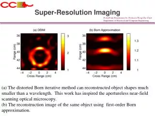
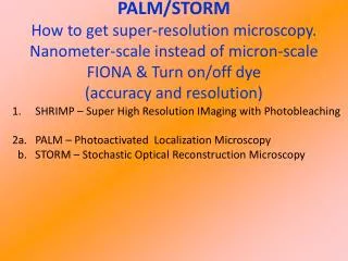
New Super-Resolution Imaging Technology for Biomedical Research CD BioSciences, a US-based biotechnology company focusing on the development of imaging technology, introduced its new services based on super-resolution imaging technology to help researchers study various cellular activities in cellular structures, such as DNA/RNA and protein interactions, DNA replication and transcription, vesicle motility, and bacterial transcription. Microscopes are the most common research tools used in biological research. Researchers typically use electron microscopes to study subcellular structures or optical microscopes to investigate whole living cells. However, traditional microscopes have limited resolution. For example, when the distance between two or more fluorophores is within a few hundred nanometers, the images become blurred. In addition, traditional microscopes cannot directly observe cellular structures and biomolecular interactions because biological structures such as protein complexes, the Golgi apparatus, and the cytoskeleton are below the diffraction limit of traditional microscopes. CD BioSciences has solved this problem with super-resolution imaging technology, which enables scientists to directly observe cell structures and biomolecular interactions, and study various cell activities in cell structure, such as DNA/RNA and protein interaction, DNA replication and transcription, vesicle movement and bacterial transcription. It has a number of advantages over traditional light microscopy, including high resolution, the ability to study protein structure at the organelle level, and the ability to follow the movement of individual molecules in cells in real time. These advantages make super-resolution imaging technology a valuable tool for researchers studying the mechanisms of cell physiology. Furthermore, super-resolution microscopy breaks the resolution barrier while maintaining the minimally invasive nature of optical microscopy. It enables the observation of fine structures and molecular activities in the 10-100 nanometer resolution range by providing 20 times the imaging resolution of optical microscopy. It works by effectively reducing the point spread function or ensuring that the fluorophores of a sample are not too close together to prevent blurring. Stimulated microscopy (SIM), stochastic optical reconstruction microscopy (STORM), and two-photon excitation microscopy (TPE) super-resolution microscopy techniques. These techniques produce super-resolution images in the low nanometer range, allowing observation of intracellular molecular interactions and fine cellular structures. emission depletion microscopy (STED), structured illumination are the most commonly used CD BioSciences specializes in the development and application of super-resolution
imaging technology. It offers a wide range of services including image analysis and consulting. For more information about the new super-resolution imaging technology, please visit https://www.bioimagingtech.com/super-resolution-imaging-technology.html. About CD BioSciences CD BioSciences is a biotechnology company committed to the development of imaging technology for many years. Its scientists can utilize high-content imaging, nanoparticle imaging, imaging flow cytometry, time-lapse imaging, and other techniques to image cell structure, cell migration, cell proliferation, pathogen infection mechanisms, and interactions between protein molecules.