Download

1 / 42
450 likes | 570 Views
Lecture 11 clinical practice. Laboratory techniques commonly used in immunology: I. Dr. Dalia Galal. Antigen -Antibody Reactions. Antigen – antibody reactions are performed to determine the presence of either the antigen or antibody. ( serological tests ).

E N D
Lecture 11 clinical practice Laboratory techniques commonly used in immunology: I Dr. Dalia Galal
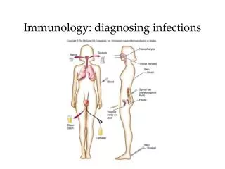
Antigen -Antibody Reactions • Antigen – antibody reactions are performed to determine the presence of either the antigen or antibody. ( serological tests ). • e.g. with a known antigen, such as influenza virus , a test can determine whether antibody to the virus is present or not . Types of serological Tests Exp. 1 Agglutination Reaction Exp. 2 Precipitation Reaction Exp. 3 Radioimmunoassay Exp. 4 Complement Fixation Exp. 5 immunofluorescence (IFA) Monoclonal antibodies
Agglutination: In this test the antigen is particulate (e.g. bacteria and red blood cells) or an inert particle (latex beads) coated with antigen. Antibody is divalent and cross links the multivalent antigen to form a lattice network or clumps (agglutination). This reaction can be performed in a tube or on a glass slide e.g. ABO blood grouping. Isolate Abs from patient Exp. 1 Agglutination Reaction
Contains patient’Abs Agglutination
Contains Abs for the specific pathogen Agglutination
Haemaggultination Tests: • Viruses can clump red blood cells from one species or another (active hemagglutination) . • This can be inhibited by specific anti-viral antibodies. • Red cells can also absorb many antigens and when mixed with specific antibodies will form clumps (passive hemagglutination) i.e. red cells are passive carriers .
Exp. 2 Precipitation Reaction • In this test antigen is in soluble form (solution). • Antibody cross -links antigen molecules to form aggregates (precipitates) in the zone of equivalence: optimal proportion of antigen and antibody. • Precipitation test can be performed in solution or in semi- solid medium (agar).
Precipitation in Agar • Single radial immunodiffusion: • Antibody is incorporated into agar and antigen introduced into the well. • As antigen diffuses into agar precipitation rings form depending on the concentration of the antigen. • Radial Immunodiffusion is used to measure IgG, IgM and complement components.
Double immunodiffusion • Antigen and antibody are placed in different wells in agar and allowed to diffuse and form precipitation lines at the points of optimal concentrations. • This method is used to determine whether antigens are related, identical or non –identical.
Exp. 4 complement Fixation • Based on the principle that antigen and antibody reaction activates complement . • Antigen and antibody, one known and the other unknown are mixed. • A measured amount of complement is added . • If antigen-antibody reaction has occurred it will combine “fix” complement. • An indicator system consisting of “sensitized” red blood cells (red blood cells plus anti-red blood cell antibody) is added. • If the complement was fixed because of antigen antibody reaction red cells will not be hemolyzed i.e. the test is positive. • If the antigen antibody reaction did not occur in the first step complement will not be fixed and will be available to lyse RBCs – a negative test.
Exp. 5 Immunofluorescence (IFA) Principle of the Test • For the determination of autoantibodies against infectious agents, cells, tissue sections. • Immunofluorescence is the labeling of antibodies or antigens with fluorescent dyes. • Fluorescein is a dye (e.g. fluorescein and rhodamine) which can be covalently attached to antibody molecules and emits greenish or red fluorescence by ultraviolet (UV) light in a fluorescent microscope. • Such labeled antibody can be used to identify antigens on surface of microorganisms ( e.g. treponemes), in histological section or in other specimens.
There are two ways of doing IF staining • Direct immunofluorescence • Indirect immunofluorescence Direct immunofluorescence • Target protein (Antigen or Ag) is fixed on the slide • Fluorescein labeled antibodies (Ab) are layered over it • Slide is washed to remove unattached Ab’s • Examined under UV light in an fluorescent microscope • The site where the Ab attaches to its specific Ag will show apple green fluorescence • Use: Direct detection of Pathogens or their Ag’s in tissues or in pathological samples
The unlabelled antibody is applied directly to the tissue substrate • Treated with a fluorochrome-conjugated anti-immunoglobulin serum • Antibody of interest if present will remain attached and can be detected by addition of fluorescent dye labeled antibody under UV light. • Advantage of indirect over direct IF • Because several fluorescent anti-immunoglobulins can bind to each antibody present in the first layer, the fluorescence is brighter than the direct test. Indirect Immunofluoresence involves a two stage process:
ANA (antinuclear antibodies) – are autoantibodies against various antigens of cell nuclei and found in patients with autoimmune diseases such as systemic lupus erythematosus (SLE). Examples of immunofluorescence assay for diagnosis of Autoimmune diseases:
ANCA (anti-neutrophil cytoplasm antibody) detects autoantibodies against neutrophil specific antigens: • cANCA (cytoplasmic) is highly specific for vasculitis. • pANCA (perinuclear) is present in a high proportion of cases of glomerulonephritis pANCA Pattern C-ANCA Pattern
Monoclonal Antibodies (Mab) • Single specificity antibodies formed by fusing a mouse B cell with a myeloma cell • A malignant tumor formed by the B cells of the bone marrow • Used in diagnosis of disease, identification of microbes and therapy. • Hybridoma: it is hybrid cell by fusing an antibody -producing B cell with a myeloma (B cell cancer)
Steps for prepration of Monoclonal Antibodies Immunize animal Harvest spleen Fuse B & myeloma Hybridoma Screen hybridomas for Abs directed against antigen of interest Hybridomas produce antibody that recognize single epitope Produce uniform, highly specific Ab in large supply
Lecture 12: clinical practice Laboratory techniques commonly used in immunology (II) Dr. Dalia Galal
Objectives: Exp. 6 Enzyme-Linked Immunosorbent Assay Exp. 7 Western Blot Exp. 8 Techniques of HLA Typing Test Exp. 9 Measurement of Phagocytosis by Phagocytes Exp. 10 Detection of T Lymphocytes Subgroup
Exp. 6 Enzyme-Linked Immunosorbent Assay (ELISA) Principle • The principle of enzyme-linked immunosorbent assay (ELISA) is based on the high specificity of antigen-antibody reactions. • ELISA may be quantified by determining the rate at which the enzyme converts a clear substrate to a colored product. • ELISA is now widely used in many areas in the medicines. • There are many different approaches to ELISA (Fig.4-1); They are typically performed as a direct or an indirect method, but can also be performed as a two antibodies sandwich method and a competitive assay.
Fig. 4-1 Principles of enzyme-linked immunosorbent assay (ELISA)
Application ELISA is a very sensitive laboratory method and can be quantitative when used in conjunction with standard curves. It can quantify the amount of antigen or antibody present in a given sample. Purpose Double Abs sandwich ELISA can be used in detecting HBsAg in serum. Attention 1. Samples and test kit need to be used under room temperature 2. Shake reagents before using 3. Slowly draw and discharge samples with micropippettor 4. Change tips between solutions 5. Filter paper and tape can’t be reused 6. Wash wells carefully to prevent overflow 7. Plate needs to be read within 10 minutes after adding the termination solution
Exp. 7 Western Blot • Protein mixture treated with detergent SDS. • Separated by sodium dodecyl sulfate Polyacrylamide gel electrophoresis SDS PAGE- lower molecular weight migrate faster. • Proteins blotted from gel to nitrocellulose membrane under electric current. • add antiserum, then secondary labelled antibody with enzyme, then subsrate
Exp. 8 Techniques of HLA Typing Test Organ and tissue transplant is necessary when an individual has suffered from a disease or an injury. HLA Typing Methods, Techniques and Results HLA typing stands for Human Leukocyte Antigen typing. The HLA antigen is present on the surface of white blood cells. White blood cells form one of the key agents of the human immune system. The HLA typing methods are used to determine whether an individual's body will accept or reject foreign tissue. The human body uses the HLA typing of the tissue to determine whether it is foreign tissue or not.
When the body rejects the transplanted organ or tissue, it may even begin to attack it. This attack leads to the area being damaged. The rejection of heart and liver tissue will almost certainly lead to the death of an individual. One of the HLA typing techniques used is a simple blood test. In addition, blood type has been classified, it will be clear whether the doctor can go ahead with the transplant procedure or not.
Exp. 9 Measurement of Phagocytosis by Phagocytes Principle This protocol describes a simple assay to measure the ability of phagocytes to bind and internalize or phagocytose bacteria (Fig.7-1). Bacteria and cells are mixed in suspension and rotated to give optimal condition for interaction. Extracellular bacteria are then removed by washing or centrifugation in sucrose solution. The amount of phagocytosis is determined by examining the stained cells under oil immersion microscope (Fig. 7-2). Application Determine the phagocytosis of macrophages and neutrophilic granulocytes and evaluate the status of innate immunity of an individual. Results Quantitate phagocytosis under oil-immersion microscopy (100×), examining at least 200 cells and counting the number of internalized bacteria in each one.
Attentions 1. Vortexing is important so that the bacteria are mixed to the single cells. For this assay, a ratio of ten bacteria to one macrophage is used to allow easy visualization and quantitation of phagocytosis. However, bacteria are well phagocytosed and readily detectable at 1:1 ratio. 2. Time periods of the tubes on a shaker and rotate end-over-end >30 min should not be used, because significant killing of many bacteria can occur quite rapidly and dead bacteria degraded by extensive exposure to 37℃ may fail to be stained and detected as phagocytosed.
Fig.7-l Mechanism of phagocytosis Fig.7-2 Phagocyte
Exp. 10 Detection of T Lymphocytes Subgroup with the APAAP Principle T lymphocyte is one of the most functionally important cells in human immune system. According to the CD (Cluster Differentiation) molecules expressed on its surface, T cell is divided into two groups, CD3+CD4+CD8 - T cell and CD3+CD4-CD8+T cell. APAAP complex is consisted of alkaline phosphatase and mouse anti-alkaline phosphatase antibody. Samples (peripheral blood mononuclear cell, PBMC) are fixed on slides, followed by the addition of primary antibody (mouse anti-human T cell CD3 McAb). Bound antibody is detected by secondary/bridge antibody (rabbit anti-mouse IgG). In this bridge antibody, one Fab is bound to primary Ab, the other Fab is bound to APAAP complex. Afterwards, the substrate solution is added to the reaction solution to demonstrate color reaction. Thus T cell subgroups expressing corresponding CD molecules are stained with the pigment (Fig. 6-1)
Application The APAAP method is employed to detect T cell subgroups in order to determine the status of patient immune functions, to diagnose some related diseases. Forexample, CD4/CD8 ratio is decreased in AIDS patients. It also used other cell markers, such as MHC-I, MHC-II antigens. Results Examine slides using 40 × microscope. Count 200 PBMC to determine percentage of positive cells. CD (+) cell: the cell nucleus stained blue, the cell membrane/plasma stained red. CD (-) cell: the cell nucleus stained blue, no color or transparent on cell membrane/plasma.