Download
1 / 40

E N D
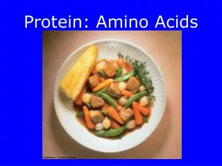
M.Prasad Naidu MSc Medical Biochemistry, Ph.D,. NON PROTEIN AMINOACIDS
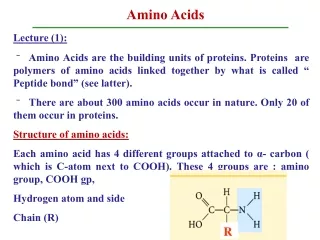
Non protein amino acids • These amino acids, although never found in proteins, perform several biological important function. • These NPAAs and D-AA speculated to be related to auto immune disease and to aging
understand the role of NPAAs and D-AA s in auto immune disease and aging, the determination of these NPAAs and D-AA s is required. • any nonprotein amino acid can be chemically incorporated into peptides, provided that appropriate methods are designed for protecting the functional group.
Nonprotein amino acids with no cytotoxicity have been known to be incorporated into proteins. For examples, • tyrosine and tryptophan residues in some proteins have been substituted with m-fluorotyrosine and 4-fluorotryptophan respectively, without any effects on the protein functions, and the 19F nuclei have been used as magnetic resonance
One NPA that has received some attention is canavanine, (L-2-amino-4-(guanidinooxy)butyric acid), the guanidinooxy structural analogue of arginine. These non protien amino acids are classified as alpha and non alpha amino acids Alpha amino acids: a) ornithine b) citruline c) arginosuccinic acid d) thyroxine e) triodothyroxine f) S-Adenosylmethionine g) Homocysteine h) 3,4-Dihydroxy phenylalanine ( DOPA) I ) creatinine j) ovathiol k) Azaserine
2) NON ALPHA amino acids: a) beta –alanine b) beta – aminoisobutyric acid c) gama – aminobutyric acid(GABA) d) aminolevulinic acid (ALA) e) taurine
Alpha - aminoacids 1) ornithine : • ornithine is precursors of polyamine Hydrolytic cleavage of the guanidino group of arginine, catalyzed by liver arginase, releases urea . The other product, ornithine, reenters liver mitochondria and participates in additional rounds of urea synthesis. Ornithine and lysine are potent inhibitors of arginase, and compete with arginine. Arginine also serves as the precursor of the potent muscle relaxant nitric oxide (NO) in a Ca2+-dependent reaction catalyzed by NO synthase 2) Citrulline : Citrulline is intermediates in the biosynthesis of urea
L-Ornithinetranscarbamoylase catalyzes transfer of the carbamoyl group of carbamoyl phosphate to ornithine,formingcitrulline & orthophosphate While the reaction occurs in the mitochondrial matrix, both the formation of ornithine and the subsequent metabolism of citrulline take place in the cytosol. • Entry of ornithine into mitochondria and exodus of citrulline from mitochondria therefore involve mitochondrial inner membrane transport systems
3)Arginosuccinic acid : • Arginosuccinic acid is intermediates in the biosynthesis of urea • Argininosuccinatesynthetaselinks aspartate and citrulline via the amino group of aspartate and provides the second nitrogen of urea. • The reaction requires ATP and involves intermediate formation of citrullyl-AMP. Subsequent displacement of AMP by aspartate then forms arginosuccinate.
In addition to patients that lack detectable argininosuccinatesynthetase activity a 25-fold elevated Km for citrulline has been reported. In the resulting citrullinemia, plasma and cerebrospinal fluid citrulline levels are elevated, and 1–2 g of citrulline are excreted daily.
4) Thyrosine and triodothyroxine : • Tyrosine forms norepinephrine and epinephrine, and following iodination the thyroid hormones triiodothyronine and thyroxine. • Use of measurement of blood thyroxine or thyroid-stimulating hormone (TSH) in the neonatal diagnosis of congenital hypothyroidism. • The amino acid tyrosine is the starting point in the synthesis of the catecholamines and of the thyroid hormones tetraiodothyronine (thyroxine; T4 ) and triiodothyronine (T3)
thioredoxinreductase, glutathione peroxidase, and the deiodinase that converts thyroxine to triiodothyronine. • The clinical history, physical examination, and lab results were all consistent with primary hypothyroidism. Accordingly, the patient was started on a low dose of thyroxine (T4 ). • It is important to begin therapy with a small dose of T4, as larger doses can precipitate serious cardiac events, due to the changes in metabolism caused by administration of the hormone. • Thyroxine (T4), free: 4.0 pmol/L (normal 10.3–21.9 pmol/L)
5)S-Adenosylmethininehomocysteine: S-Adenosylmethionine, the principal source of methyl groups in metabolism, contributes its carbon skeleton to the biosynthesis of the polyamines spermine and spermidine. Homocystinuria HomocystinuriaCystathionine -synthase Lens dislocation, thrombotic vascular disease, mental retardation, osteoporosis AR Homocystinuria 5,10-Methylenetetrahydrofolate reductase Mental retardation, gait and psychiatric abnormalities, recurrent strokes ,Mental retardation, hypotonia, seizures, megaloblastic anemia
Pathways, enzymes, and coenzymes involved in the homocystinurias.Methionine transfers a methyl group during its conversion to homocysteine. • Defects in methyl transfer or in the subsequent metabolism of homocysteine by the pyridoxal phosphate (vitamin B6)-dependent cystathionine b-synthase increase plasma methionine levels. • Homocysteine is transformed into methionine via remethylation. This occurs through methioninesynthase, a reaction requiring methylcobalamin and folic acid. • Deficiencies in these enzymes or lack of cofactors is associated with decreased or normal methionine levels. In an alternative pathway, homocysteine can be remethylated by betaine:homocysteine methyl transferase
Life-threatening vascular complications (affecting coronary, renal, and cerebral arteries) can occur during the first decade of life and are the major cause of morbidity and mortality. • Classic homocystinuria can be diagnosed with analysis of plasma amino acids, showing elevated methionine and presence of free homocystine.
Total plasma homocysteine is also extremely elevated (usually >100 M). Treatment consists of a special diet restricted in protein and methionine and supplemented with cystine. • In approximately half of patients, oral pyridoxine (25–500 mg/d) produces a decrease in plasma methionine and homocystine concentration in body fluids. • Folate and vitamin B12 deficiency should be prevented by adequate supplementation. Betaine is also effective in reducing homocystine levels.
6) 3-4 Dihydrophenylalanine(DOPA): • Neural cells convert tyrosine to epinephrine and norepinephrine. While dopa is also an intermediate in the formation of melanin, different enzymes hydroxylate tyrosine in melanocytes. • Dopadecarboxylase, a pyridoxal phosphate-dependent enzyme, forms dopamine. Subsequent hydroxylation by dopamine -oxidase then forms norepinephrine. • In the adrenal medulla, phenylethanolamine-N-methyltransferase utilizes S-adenosylmethionine to methylate the primary amine of norepinephrine, forming epinephrine . • Tyrosine is also a precursor of triiodothyronine and thyroxine. • DOPA ... related to dopaminerelationship to Parkinson's Disease
Dopamine, Norepinephrine, and Epinephrine 1. SYNTHESIS OF THE CATECHOLAMINE NEUROTRANSMITTERS • These three neurotransmitters are synthesized in a common pathway from the amino acid L-tyrosine. • The first and rate-limiting step in the synthesis of these neurotransmitters from tyrosine is the hydroxylation of the tyrosine ring by tyrosine hydroxylase, a tetrahydrobiopterin(BH4)-requiring enzyme. The product formed is dihydroxyphenylalanine or DOPA. • The phenyl ring with two adjacent OH groups is a catechol, andhence dopamine, norepinephrine, and epinephrine are called catecholamines.
The second step in catecholamine synthesis is the decarboxylation of DOPA to form dopamine. This reaction, like many decarboxylation reactions of amino acids,equirespyridoxal phosphate. • Dopaminergic neurons (neurons using dopamine as a neurotransmitter) stop the synthesis at this point, because these neurons do not synthesize • the enzymes required for the subsequent steps. Neurons that secrete norepinephrine synthesize it from dopamine in a hydroxylation reaction catalyzed by dopamine -hydroxylase (DBH). This enzyme is present only within the storage vesicles of these cell • Although the adrenal medulla is the major site of epinephrine synthesis, it is also synthesized in a few neurons that use epinephrine as a neurotransmitter.
7)Creatinine : Creatinine is formed in muscle from creatine phosphate by irreversible, nonenzymatic dehydration and loss of phosphate. Since the 24-h urinary excretion of creatinine is proportionate to muscle mass, it provides a measure of whether a complete 24-h urine specimen has been collected. • Glycine, arginine, and methionine all participate in creatine biosynthesis. Synthesis of creatine is completed by methylation of guanidoacetate by S-adenosylmethionine.
Normal values : • Creatinine : 200 mol/L (44–80 mol/L) • Female -- 44–80 mol/L 0.5–0.9 ng/mL • male -- 53–106 mol/L 0.6–1.2 ng/mL
8) Ovathiol:- • Sulfur containing amino acid found in fertilized Eggs, and acts as an antioxidant 9) Azaserine: (antibiotic) Purine deficiency states, while rare in humans, generally reflect a deficiency of folic acid. Compounds that inhibit formation of tetrahydrofolates and therefore block purine synthesis have been used in cancer chemotherapy. Inhibitory compounds and the reactions they inhibit include azaserine, diazanorleucine, 6-mercaptopurine , and mycophenolic acid .
II) Non--Amino Acids: • Non--amino acids present in tissues in a free form include -alanine, -aminoisobutyrate, and -aminobutyrate (GABA). -Alanine is also present in combined form in coenzyme A and in the -alanyldipeptidescarnosine, anserine and homocarnosine . • 1) Beta-Alanine & -Aminoisobutyrate: • Alanine and -aminoisobutyrate are formed during catabolism of the pyrimidinesuracil and thymine, respectively . Traces of -alanine also result from the hydrolysis of -alanyldipeptides by the enzyme carnosinase. -Aminoisobutyrate also arises by transamination of methylmalonatesemialdehyde, a catabolite of L-valine . • The initial reaction of -alanine catabolism is transamination to malonatesemialdehyde. Subsequent transfer of coenzyme A from succinyl-CoA forms malonyl-CoAsemialdehyde, which is then oxidized to malonyl-CoA and decarboxylated to the amphibolic intermediate acetyl-CoA.
Analogous reactions characterize the catabolism of -aminoisobutyrate. Transamination forms methylmalonatesemialdehyde, which is converted to the amphibolic intermediate succinyl-CoA by reactions 8V and 9V of. Disorders of -alanine and -aminoisobutyrate metabolism arise from defects in enzymes of the pyrimidine catabolic pathway. Principal among these are disorders that result from a total or partial deficiency of dihydropyrimidinedehydrogenase.
2) beta-AlanylDipeptides : • The -alanyldipeptidescarnosine and anserine (N -methylcarnosine) activate myosin ATPase, chelate copper, and enhance copper uptake. -Alanyl-imidazole buffers the pH of anaerobically contracting skeletal muscle. • Biosynthesis of carnosine is catalyzed by carnosinesynthetase in a two-stage reaction that involves initial formation of an enzyme-bound acyl-adenylate of -alanine and subsequent transfer of the -alanyl moiety to L-histidine.
Hydrolysis of carnosine to -alanine and L -histidine is catalyzed by carnosinase. The heritable disorder carnosinase deficiency is characterized by carnosinuria. • Homocarnosine, present in human brain at higher levels than carnosine, is synthesized in brain tissue by carnosinesynthetase. Serum carnosinase does not hydrolyze homocarnosine. Homocarnosinosis, a rare genetic disorder, is associated with progressive spastic paraplegia and mental retardation.
3) gama-Aminobutyrate • gama-Aminobutyrate (GABA) functions in brain tissue as an inhibitory neurotransmitter by altering transmembrane potential differences. • GABA is formed by decarboxylation of glutamate by L -glutamate decarboxylase. Transamination of -aminobutyrate forms succinatesemialdehyde, which can be reduced to -hydroxybutyrate by L -lactate dehydrogenase, or be oxidized to succinate and thence via the citric acid cycle to CO2 and H2O.
A rare genetic disorder of GABA metabolism involves a defective GABA aminotransferase, an enzyme that participates in the catabolism of GABA subsequent to its postsynaptic release in brain tissue. • Defects in succinicsemialdehydedehydrogenase are responsible for another rare metabolic disorder of -aminobutyrate catabolism characterized by 4-hydroxybutyric aciduria.
4) amino levulinic acid (ALA) : • ALA is intermediate in the synthesis of porphyrin (finally heme)
5) Taurine : • Taurine (2-aminoethylsulphonic acid) is a non-protein aminoacid present in almost all animal tissues and the most abundant free intracellular aminoacid in human cells. • In humans, it is considered to be a “semi-essential aminoacid” since it can be synthesized from other sulfonicaminoacids such as methionine and cysteine, in the presence of vitamin B6,2,3 but endogenous production is insufficient, so that it needs to be provided through diet. Taurine is found in association with bile acids.
Biological effects of taurine in the context of diabetes • Biological EffectMechanismofTaurine • Antioxidant action By inhibiting ROS generation at mitochondria Osmoregulation By counteracting osmotic inbalance through cellular membrane due • to hyperglycaemia • Antiinflammatory effects By interfering the formation of inflammatory mediators Glucose Homeostasis By interfering the insulin signalling pathway acting upon UCP2 protein
Estimation of NPAAs : 1) The aim of our study was to analyze NPAAs and D-AA s in biosamples by means of capillary electrochromatogrphy (CEC) using a chiralpracticle- lodedmonolithiac column with flurrosense detection for high sensitivity. 2) capillary electrophoresis 3) High perfromance liquid chromatography(HPLC) 4) laser-induced flurosecne (LIF) 5) scanning electro microscopy (SEM)
Location of the regions of ordered secondary structures for b-residues in f–q–y space. The a-helix and b-sheet are the classical structures for poly a-amino acids. b-residues occurring in the appropriate shaded region can be accommodated without disruption of secondary structures. The 12-helix and 14-helix are well characterised secondary structures for poly b-peptides.