Download
1 / 40
400 likes | 404 Views
Part II: Genetics – The basis of heredity. FACT: Related individuals resemble each other more than randomly chosen individuals do Within populations Among populations within species Within species among species Etc. …WHY??. Resemblance among relatives…”heredity”.

E N D
Part II: Genetics – The basis of heredity • FACT: Related individuals resemble each other more than randomly chosen individuals do • Within populations • Among populations within species • Within species among species • Etc. …WHY??
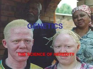
Resemblance among relatives…”heredity” • Information is passed from parent to offspring • Information is passed largely intact • That information encodes the phenotype • Phenotype is any property of an organism that can be attributed to that organism (height, weight, eye color, obnoxiousness, etc.)
Outline of today’s lecture – (Ch. 12) • Review of (some) Eukaryotic cell structures • Schematic overview of mitosis • The mitotic spindle • Cytokinesis • Cell cycle control
Review of Eukaryotic cell structures • Nucleus • Surrounded by membrane • Contains the genetic material (DNA) • During interphase, DNA uncoiled, complexed with proteins, called “chromatin” • In mitosis, condenses into chromosomes
Review of Eukaryotic cell structures • Nucleolus • Point of synthesis of ribosomal RNA • Ribosomal subunits assembled Nucleolus
Review of Eukaryotic cell structures • Centrosomes • Region near nucleus that is the cellular “microtubule organizing center” • Associated with formation of spindle fibers • Form “spindle poles” during mitosis
Review of Eukaryotic cell structures • Cytoskeleton • Provides mechanical support to the cell • Dynamic (disassemble and reassemble) • Constructed of microtubules, microfilaments, intermediate filaments
Microtubules: Structure and Function • Composed of polymers of dimers of α-tubulin, β-tubulin • Lengthen by adding dimers, shorten by losing dimers • “Molecular motors” can move along microtubules
Mitosis and the Cell Cycle • All organisms must grow and reproduce • During organismal growth, all cells must grow and divide • Genetic information must be faithfully passed between parent cell and daughter cells • MITOSIS is the process by which genetic information is passed from parent to daughter cells
Some notation • Chromosome • Centromere • Spindle pole (centrosome) • Spindle fiber • Nuclear envelope
F M G1 (Interphase) • Each chromosome is a single, unreplicated double strand of DNA • One chromosome from each parent (Male, Female) forms a Homologous Pair (= “homologs”) • Chromosomes in nucleus, surrounded by nuclear membrane • Single centrosome
F F M M S-phase (DNA synthesis) • After DNA replication, TWO “sister chromatids” are present for each homolog • Each sister chromatid is the SAME double-stranded DNA molecule • Attached by proteins, tightly at centromere and more loosely throughout their length
F F M M G2 (Interphase) • Duplication of centrosomes • Cell receives signal to enter mitosis
F F M M M1 (Early Prophase) • Chromosomes condense • Mitotic spindle forms from centrosome • Centrosomes begin to migrate to poles of cell • Nucleoli disappear
F F M M M2 (Mid-prophase) • Chromosomes fully condensed • Centrosomes complete migration to the poles • Nuclear envelope begins to degrade • Spindle fibers enter nuclear area from the pole • Kinetochores form at centromeres
F F M M M3 (Prometaphase) • Nuclear envelope completely degraded • Kinetochores form at centromeres • Some spindle fibers attach at kinetochores • Sister chromatids attached to opposite poles
F F M M M4 (Metaphase) • Spindle fibers attached to centromere at kinetochore • All sister chromatids attached to opposite poles • Chromosomes migrate to center plane of cell, “metaphase plate”
M5 (Early Anaphase) • Protein bond between sister chromatids degrades • Sister chromatids separate, begin migration toward opposite poles • Poles move farther apart as non-kinetochore spindle fibers lengthen
M5 (Late Anaphase) • Chromosomes (no longer “chromatids”) have reached poles
M6 (Telophase) • Non-kinetochore spindle fibers continue to elongate cell • Nuclear envelopes begin to form at poles • Chromosomes de-condense back into chromatin • Nucleoli re-form, cytokinesis begins
The mitotic spindle: Structure and Function • Made of microtubules, associated proteins • Cytoskeleton partially disassembles to provide materials • Assembly starts in centrosomes • Centrosomes “pushed” away from each other as microtubules grow
The mitotic spindle: Structure and Function • Some spindle fibers grow and attach to the kinetochore while sister chromatids are still attached • “Equilibrium” reached when sister chromatids are midway between the poles (metaphase plate)
Microtubules: Structure and Function • Composed of polymers of dimers of α-tubulin, β-tubulin • Lengthen by adding dimers, shorten by losing dimers • “Molecular motors” can move along microtubules
The mitotic spindle: Structure and Function • Hypothesized mechanism for chromosome movement
The mitotic spindle: Structure and Function • Experimental Test of the hypothesized mechanism for chromosome movement • What is an obvious alternative hypothesis?
Cytokinesis – Animal cells • Cleavage furrow formed by contraction of a ring of microfilaments • Associated with actin and myosin (motor protein system) • Forms along metaphase plate
Cytokinesis – Plant cells • Plants have cell walls • Vesicles (derived from Golgi) move along microtubules to the middle of the cell • Vesicles coalesce to form a “cell plate”; membrane derived from vesicles • Cell plate membrane fuses with outer cell membrane, forms daughter cells
Cell Division in Prokaryotes - Binary fission • Prokaryotes preceded Eukaryotes by billions of years • Bacterial chromosome is circular DNA molecule • Origin of replication
Cell Division in Prokaryotes - Binary fission • As DNA replication proceeds, one copy of the origin migrates to each end of the cell
Cell Division in Prokaryotes - Binary fission • Cell membrane grows inward • New cell wall forms • Two daughter cells result • Migration of origin(s) is similar to migration of centromeres
Cell cycle - Regulation • Timing and rate of cell division are critical • Chemical signals regulate “checkpoints” in the cell cycle • RESULTS of certain chemical processes are “checked” • If results not appropriate, cell division does not proceed • Evidence from cell fusion experiments
Cell cycle - Regulation • G1 checkpoint: Cell will not enter into S phase unless appropriate signal is received • Many cells in adult mammals are in G0 and thus do not divide (e.g., nerve) • If not, enters non-dividing “G0” phase • Variety of external chemical cues to trigger entry into G1 and G2 phase (or not)
Cell cycle - Regulation • Additional Regulatory Checkpoints • Cell will not proceed through S-phase unless DNA synthesis completed • M-checkpoint: Cell will not enter anaphase until chromosomes lined up on metaphase plate
Cell cycle - Regulation • Cell cycle controlled by two types of proteins, kinases and cyclins • Kinases active when attached to cyclins • E.g., “Maturation-promoting factor” MPF
Cell cycle regulation – internal and external controls • M-checkpoint: if kinetochores not attached to spindle microtubules, sister chromatids remain attached • G1-checkpoint: growth factors / receptors • E.g., platelet-derived gf (PDGF), healing wounds • Density-dependent inhibition (nutrient concentration below a certain level) • Anchorage-dependence
For Wednesday: Ch. 13 • Meiosis • Sexual Life Cycles