Download
1 / 63
650 likes | 684 Views
Malaria. By Prof. Inas Zakaria Ahmed & Dr. Ahmad Hassaneen. Objectives. Knowledge of Malaria Disease Organism, pathogenesis, diagnostics, treatment, prevention and epidemiology. The Organisms. Four species of Plasmodium cause malaria in humans: Plasmodium falciparum,

E N D
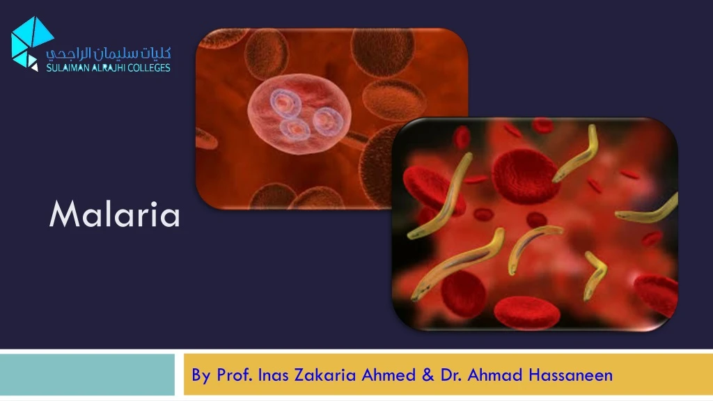
Malaria By Prof. Inas Zakaria Ahmed & Dr. Ahmad Hassaneen
Objectives • Knowledge of Malaria Disease • Organism, pathogenesis, diagnostics, treatment, prevention • and epidemiology
The Organisms • Four species of Plasmodium cause malaria in humans: • Plasmodium falciparum, • Plasmodium vivax, • Plasmodium malariae, & • Plasmodium ovale. • A fifth species, P. knowlesi, a pathogen of monkeys, recently been recognized to cause severe disease in humans in Asia. • The two most common species are Plasmodiumvivax & Plasmodium falciparum, with falciparum being the most pathogenic of all. Quoted Benjamin Wielgosz,
Plasmodium species Plasmodium malariaeis much less common than falciparum and vivax Plasmodium ovaleis rare except in West Africa, where it seems to replace P. vivax.
How serious is the problem ? - Half of the world’s population is at risk for malaria. - Almost 250 million new cases are estimated to occur each year. - In 2015, malaria caused 212 million cases of malaria & 429 000 deaths. - In 2015, 91 countries had ongoing malaria transmission. - In 2015, Sub-Saharan Africa was home to 90% of malaria cases & 92% of malaria deaths.
How serious is the problem ? ■Malaria today is generally limited to the tropics & subtropics, although outbreaks in Turkey prove the capacity of this disease to reappear in areas cleared of this disease. ■ Malaria in moderate zones is relatively uncommon, although severe epidemic outbreaks may occur when the largely non-immune populations of these areas are exposed; it is usually temporary & relatively easy to control or eradicate. ■ Tropical malaria shows more stable transmission and difficult to control. eradicate. ■ Malaria generally disappears at altitudes above 6000 ft.
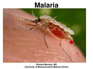
Epidemiology Malaria ■ The usual route of infection is through the bite of an infected female Anopheles mosquito. ■ All forms of malaria can be transmitted also by the following : 1-Transplacentally 2-Blood transfusion 3-Needles shared among drug abusers. ►Such cases do not develop a liver infection ; thus, relapse does not occur.
Natural Transmission & Life Cycle • - Transmission causing human infection results from bite of an infected female Anopheles mosquito, through which the sporozoites injected into the bloodstream. • - Sporozoites rapidly (within 1 hr) enter parenchymal cells of the liver, where first stage of development in humans takes place (exo-erythrocytic phase). • - Subsequently, numerous asexual progeny, the merozoites ,are released from liver cells after rupture of schizonts, enter the bloodstream, & invade erythrocytes. • - The merozoites do not return from red blood cells to liver cells. • N.B. P. vivax andP. ovaleproduce a latent form (hypnozoite) in the liver; this form is the cause of relapses seen with vivax and ovale malaria.
I=Infective stage D=Diagnostic stage
Natural Transmission & Life Cycle - Parasites in red cells multiply in a species-characteristic fashion, breaking out of their host cells synchronously. - This is the erythrocytic cycle, with successive broods of merozoites appearing at 48-hour intervals (P vivax, P falciparum, & P ovale) or every 72 hours (P malariae) - During erythrocytic cycles, certain merozoites become differentiated as male or female gametocytes.
Malaria Life cycle ●Merozoites released from liver cells infect red blood cells & differentiates into a ring-shaped trophozoite. ● Then grows into an ameboid form & differentiates into a schizont filled with merozoites. ● Released merozoites infect new erythrocytes. ● This cycle in RBCs repeats at regular intervals typical for each species, causing typical recurrent symptoms of chills, fever, & sweats in malaria patients. ● In human RBCs some merozoites develop into male & female gametocytes. These gametocyte-containing RBCs are ingested by female mosquito & within her gut, produce female macrogamete and sperm like male microgametes.
Clinical picture As disease progresses , splenomegaly &, to a lesser extent, hepatomegaly appear. A normocytic anemia develops , particularly in P falciparum infections. Untreated infection by P. falciparum is potentially life-threatening as a result of extensive brain damage (cerebral malaria) and kidney damage (black water fever). Malaria by the other three plasmodia is usually self-limited, with a low mortality rate. But, relapses of P. vivax & P. ovale malaria can occur up to several years after infection.
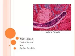
Malaria The Malaria parasites (Plasmodium species) infect both blood and tissues. (multi-organ disease)
Malaria Paroxysm (attack) Differs in different species, paroxysms may recur at regular 48- or 72-hour intervals, P falciparum pyrexia may last 8 hours or longer & may exceed 41C. . lasting several hours, shows a spiking fever that may reaches 40 C or more. During which parasites invade new red cells. Ends the episode, fever subsides, & patient falls asleep & later awakes feeling relatively well. lasting from 15 min. -1 hour, Nausea, vomiting, & headache are common
Malaria Relapse & Recrudescence Plasmodium vivax & P ovale may persist as dormant forms, or hypnozoites in liver , after parasites have disappeared from the peripheral blood, after resolution of the primary infection. Reappearance of an erythrocytic infection (Relapse) occurs when merozoites from hypnozoites in liver cells break out, & not phagocytosed in bloodstream & succeed in re-establishing a red cell infection (clinical malaria). Without treatment, P vivax & P ovale infections may persist as periodic relapses for up to 5 years. Over 50% of P. vivax infections in Thailand relapse, whereas in India relapse proportion is nearly 20%
Malaria Relapse & Recrudescence Recrudescence is the return of symptoms after a symptom-free period. It is caused by parasites surviving in the blood as a result of inadequate or ineffective treatment. It is due to persistence of drug resistant parasite. In Falciparum : Disease appears after 2-3 weeks (may be more) of completion of treatment Diagram shows course of malaria infection after mosquito bite (sp). There is prepatent period (p) between sporozoite inoculation & detection of parasites in blood. The blue line shows microscopic threshold (ie, limit of detection) & yellow area represents a subpatent parasitaemia. The orange area represents an asymptomatic patent parasitaemia. The red line represents a clinical threshold, or parasitaemia which produces paroxysms & other clinical symptoms (pink area). Malaria - Tulane University www.tulane.edu/~wiser/protozoology/notes/malaria.html
Why P. falciparum malignant malaria ? Plasmodium vivax, P malariae, & P ovaleparasitaemia is of relatively low grade, because parasites favor either young or old red cells but not both; however P falciparum invades red cells of all ages, including the erythropoietic stem cells in bone marrow, so parasitaemia is very high. Multiple merozoites can infect a single erythrocyte → three or even four small rings may be seen in an infected cell.
Why P. falciparum malignant malaria ? ●P. falciparum causes parasitized red cells to produce numerous projecting knobs that adhere to the endothelial lining of blood vessels, with resulting obstruction, thrombosis, & local ischemia. ● Trophozoite stages & schizonts of P. falciparum rarely seen in blood films except in very heavy infections because they are sequestered in the liver & spleen. ● P. falciparum infections more serious than others, with higher rate of severe & frequently fatal complications[cerebral malaria, gastrointestinal disorders, algid malaria (shock), blackwater fever ]. Molecular Aspects of Severe Malaria. Clinical Microbiology Reviews, July 2000; ● Considering malaria in DD in patients with fever & history of travel to an endemic area is critical; because delays in therapy lead to severe illness or death with falciparum malaria.
Cytoadherence Cytoadherence mediated by several different processes. The most important is high-molecular-weight parasite-derived proteins termed P. falciparum erythrocyte membrane protein 1 (PfEMP-1). PfEMP-1 is transcribed, synthesized & stored within parasite, then exported to the surface of the infecting erythrocyte. These deposits cause humps or knobs on the surface of red cell (Figure) and these are the points of attachment to vascular endothelium. Farrar et al. Manson's Tropical Diseases, 23th edition, 2014.
ROSETTING ►Erythrocytes containing mature parasites also adhere to uninfected erythrocytes, leading to formation of ‘rosettes’ when suspensions of parasitized erythrocytes are viewed under the microscope (Figure 43.9). ►Rosetting shares some characteristics of cytoadherence. But rosetting is inhibited by certain heparin subfractions and calcium chelators, unlike cytoadherence. Figure 43.9 Rosetting. (A) Uninfected red blood cells bind to an infected erythrocyte. (A, Courtesy of RachaneeUdomsangpetch. B, Courtesy of David Ferguson.)
Plasmodium Falciparum ●P. falciparum ring stage is often seen in the host cell at the edge or periphery of cell membrane, appearing almost as if it were “stuck” on the outside of cell. This is called the appliqué or accolé position andis distinctive for this species. ● Its gametocytes are crescent-shaped (“banana-shaped”), whereas those of the other plasmodia are spherical. ● P. falciparum, similar to P. knowlesi and P. malariae, does not produce hypnozoites in the liver → no relapses . ● Multiple merozoites can infect a single erythrocyte → three or even four small rings may be seen in an infected cell.
Plasmodium Falciparum The incubation period of P. falciparum is the shortest of all the plasmodia, ranging from 7 to 14 days, and does not extend for months or years. After the early influenza like symptoms, P. falciparum rapidly produces daily (quotidian) chills and fever as well as severe nausea, vomiting, and diarrhea. The periodicity of the attacks then becomes tertian (every 48 hours), and fulminating disease develops. The term malignant tertian malaria is used for this infection.
Plasmodium Falciparum • Involvement of the brain (cerebral malaria) is most often seen in this infection. Capillary plugging due to accumulation of malarial pigment and masses of cells can result in coma and death. • Kidney damage also occurs, resulting in an illness called Black water fever. • Intravascular hemolysis with rapid destruction of RBCs produces a marked hemoglobinuria and can result in acute renal failure, tubular necrosis, nephrotic syndrome, and death.
Plasmodium Falciparum ♦ A very important feature of P. falciparum is chloroquine resistance. ♦ Chloroquine-resistant strains now predominate in most areas of the world where malaria is endemic. ♦ Chloroquine resistance is mediated by a mutation in the gene encoding the chloroquine transporter in the cell membrane of the organism.
Plasmodium vivax ▪The Duffy blood group antigen on the RBC surface is the primary receptor for P. vivax. ▪Infected RBCs are usually enlarged and contain numerous pink granules or Schüffner dots. ▪ Gametocytes are round. ▪ Incubation period (usually 10 to 17 days). ▪P. vivax causes “benign tertian malaria,”which refers to the cycle of paroxysms every 48 hours (in untreated patients) and the belief that most patients tolerate the attacks and can survive for years without treatment
Plasmodium ovale ▪ P. ovale is similar to P. vivax in many respects, including its selectivity for young flexible erythrocytes. As a consequence, the host cell becomes enlarged and distorted, usually in an oval form. ▪ Schüffner dots appear as pale pink granules, and the cell border is frequently fimbriated or ragged. ▪ The clinical picture of tertian attacks for P. ovale (benign tertian or ovale malaria) infection is similar to that for P.vivax.
Plasmodium malariae ▪ In contrast with P. vivax & P. ovale, P. malariae infect only mature RBCs with relatively rigid cell membranes. As a result, the parasite’s growth must conform to the size & shape of the RBC. • ▪ So , there is no red cell enlargement/distortion, as in P. vivax & P. ovale, but it has a distinctive shapes of the parasite in the host cell: “band and bar forms”. • ▪ Incubation period for P. malariae is the longest of plasmodia spp., (18-40 days) and may extend up to several months or years. • ▪The fever patterns is 72 hours (quartan malaria) in periodicity.
Plasmodium Knowlesi known as a parasite of long-tailed macaques (Monkeys) but not considered a significant human parasite until 2004, when large number of human infections were identified in far east.
Plasmodium Knowlesi • Transmitted by Anopheles leucosphyrusgroup of mosquitoes resides in upper shady parts of forests & has infrequent contact with humans. • Similar to P. falciparum; P. knowlesi invade young & old RBCs, which allows development of high levels of parasitaemia. • - It does not sequester in the microvasculature, and neurologic complications seen with P. falciparum infection have not been described.
Laboratory Diagnosis [1] - Malaria diagnosed by microscopic examination of the blood. Thick & thin blood films made on clean, glass slides. The thick smear is used to screen for the presence ofOrganisms, & the thin smear is used for species identification (differ in ttt). The thick film done by circular motion with the corner of the second slide. Farrar et al. Manson's Tropical Diseases, 23th edition, 2014.
Laboratory Diagnosis [2]-Antigen detection using a rapid diagnostic test (RDT): RDTs use immunochromatographic strip technology and use monoclonal antibodies directed at either species-specific or pan- Plasmodium targets. These tests are simple, rapid (results in <20 minutes), and inexpensive. • P. falciparum–specific monoclonal antibodies have been developed for histidine rich protein 2 (HRP-2) and P. falciparum lactate dehydrogenase (PLDH) and aldolase enzymes.
Laboratory Diagnosis [3]-Polymerase chain reaction (PCR)-based test for Plasmodium nucleic acids or an enzyme-linked immunosorbent assay (ELISA) test for a protein specific for P. falciparum . J. Clin. Microbiol. December 2012 vol. 50 no. 12 4012-4019
Malaria in Pregnancy ▪Pregnant women in 2nd &3rd trimesters are more likely to develop severe malaria than other adults , often complicated by pulmonary edema and hypo-glycaemia. ▪ Fetal death & premature labour are common. ▪ Postpartum bacterial infection is a common complication in these cases. ▪ Falciparum malaria has also been associated with severe mid-trimester haemolytic anaemia in Nigeria, and may requires transfusion, in addition to antimalarial treatment & folate supplementation.
Sickle cell anaemia & Malaria Racial Immunity Within a species, different races may show differences in susceptibility to infections. This is known as racial immunity. Such racial differences are known to be genetic in origin, and by selection and inbreeding. Genetic resistance to Plasmodium falciparum malaria: It is seen in some parts of Africa and the Mediterranean coast and is attributed to the hereditary abnormality of the red blood cells (sickle cell ) prevalent in the area.
Sickle cell anaemia & Malaria Michael Lanzer & his Team at Heidelberg University in Germany and the Biomedical Research Center in Burkina Faso, used powerful electron microscopy techniques to compare healthy red blood cells with : 1-Normal cells infected with Plasmodium falciparum. 2- Cells from people carrying the mutated “S” gene (sickle-cell disease) infected with Plasmodium falciparum. Mutation lead to the substitution of a single amino acid in the hemoglobin molecule, causing the haemoglobin to aggregate abnormally inside the cell → cells deform into a half-moon shape — the 'sickle cells' that give the disease its name.. Sickle Cell Disease Association of Florida . (2012). Sickle Cell Disease. Retrieved 2015, from http://scdaflorida.com/resources.html
G6PD deficiency & Malaria Glucose-6-phosphate dehydrogenase (G6PD) deficiency is an X-linked recessive disorder of the erythrocyte. G6PD deficiency is mainly found in areas where malaria is or has been endemic. In these areas, malaria is treated with drugs that can cause severe hemolysis in G6PD-deficient individuals. G6PD mutations offer protection against malaria, but no clear link had been made between these mutations and infection with malaria parasites. As G6PD deficiency leads to increased oxidative stress in red blood cells, this may in turn have a negative influence on the parasite. [Louicharoen et al. (2009) Science 326:1546-1549]
Anti-Malarial Drugs Classes of antimalarial drugs • Tissue Schizonticides: Drugs eliminate developing or dormant liver forms • Blood schizonticides: Drugs act on erythrocytic parasites are; • Gametocide: Drugs that kill sexual stages and prevent transmission to mosquitoes. • No single available agent can achieve radical cure, • (eliminate both hepatic & erythrocytic stages). • Few available agents are causal prophylactic drugs, • (capable of preventing erythrocytic infection).
Anti-Malarial Drugs Blood schizonticides Chloroquine , Amodiaquine Quinine, Mefloquine Malarone (250mg Atovaquone+ 100mg proguanil) Tissue schizonticides Primaquine Malarone (250mg Atovaquone+ 100mg proguanil) Gametocidal Primaquine Quinine
Treatment Treatment of malaria is complicated. The main criteria used for choosing specific drugs are the severity of the disease and whether the organism is resistant to chloroquine.
Treatment • Chloroquine resistance is determined by the geographical location where the infection was acquired rather than by laboratory testing. • Chloroquine is the drug of choice for treatment of uncomplicated malaria caused by P. falciparum or P. malariaein areas without chloroquine resistance. • Chloroquine kills the merozoites, thereby reducing the parasitemia, but does not affect the hypnozoites of P. vivax and P. ovale in the liver. • Hypnozoites are killed by primaquine, which must be used to prevent relapses.
Treatment Primaquine May induce severe hemolysis in those with G6PD deficiency, so testing for this enzyme should be done before the drug is given. It should not be given if the patient is severely G6PD deficient. If primaquine is not given, one approach is to wait to see whether symptoms recur and then treat with chloroquine.
Treatment Uncomplicated, chloroquine-resistant P. falciparum infection is treated with either : • Coartem (artemether plus lumefantrine) • Malarone (atovaquone and proguanil).
Treatment In severe complicated cases of chloroquine-resistant falciparum malaria, intravenous administration of either • Artemisinin derivative (e.g. artesunate or artemether) • Quinidine . • The Artemisinins are active component of a herbal medicine that has been used as an antipyretic in China for over 2000 years. Artemisinins are inexpensive, has few side effects, most plasmodia have not developed resistance to them. However, Resistance to artemisinins was reported on the Cambodia -Thailand border in 2009, & then reported in Myanmar & Vietnam.
Prevention (Chemoprophylaxis) [1]-Chloroquine is used for Chemoprophylaxis for travelers in areas where P. falciparum is sensitive to that drug. [2]-Chemoprophylaxis for travelers to areas where chloroquine-resistant P. falciparum is endemic consists of either: a- Mefloquine . b-Doxycycline. c- Malarone (a combination ofatovaquone and proguanil). [3]-Travelers to areas with other three plasmodia are found should take chloroquine starting 2 weeks before arrival in the endemic area &continuing for 6 weeks after departure. This should be followed by a 2-week course of primaquine if exposure was high. Primaquine will kill the hypnozoites of P. vivax and P. ovale.