Download

1 / 23
1.17k likes | 5.43k Views
ePoster Presentation: 2011 ISS Meeting San Diego. Posterolateral Corner of the Knee: MRI, Anatomy, Functional Considerations, and Injuries. Anthony J Peduto 1 , Tushar Singh 1 , Donald L Resnick 2 Department of Radiology, Westmead Hospital and Sydney University, Australia 1

E N D
ePoster Presentation: 2011 ISS Meeting San Diego Posterolateral Corner of the Knee: MRI, Anatomy, Functional Considerations, and Injuries Anthony J Peduto1, Tushar Singh1, Donald L Resnick2 Department of Radiology, Westmead Hospital and Sydney University, Australia1 Musculoskeletal Imaging Section, Department of Radiology, University of California, San Diego2
The Posterolateral corner (PLC) Jungle • Surgeons sometimes refer to the PLC as the dark side of the knee • The structures occupying the PLC are thought to be complex, and are poorly understood, like the darkest of jungles • Diagnosis of injuries may be difficult on clinical assessment, and many of the structures are out of clear sight of the arthroscope . • MRI offers the best opportunity for early diagnosis, but has been limited by image resolution and full understanding of the anatomy ISS 2011: PLC of the Knee; Peduto A, Singh T, Resnick D
Posterolateral corner Jungle • Much of the confusion is due to lack of agreement about: • What structures actually constitute the PLC • Lack of uniform terminology • Published diagrams show significant variability in position and naming of various structures ISS 2011: PLC of the Knee; Peduto A, Singh T, Resnick D
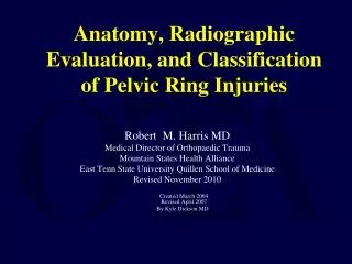
B A The red arrow on the Grays anatomy drawing (A) shows the popliteofibular ligament in its correct position and as a robust ligament (pop muscle belly is not shown). Compare this to the Netter drawing (B) which does not show the popliteofibular ligament at all, instead the V shaped arcuate ligament is incorrectly depicted as a robust structure ISS 2011: PLC of the Knee; Peduto A, Singh T, Resnick D
A B The position of the Fabello-fibular ligament is incorrectly shown positioned deep to popliteus in drawing A and is called the Fibulocolateral ligament (correctly labelled FFL in B) The FFL, Lateral colateral ligament (LCL), and biceps tendon are incorrectly shown to insert together on the fibular head at the styloid region The Arcuate ligament (AL) is incorrectly shown as an arch shaped structure in both diagrams ISS 2011: PLC of the Knee; Peduto A, Singh T, Resnick D
How should we group the structures of the PLC? Seebacher JR, J Bone Joint Surg Am 64:536-541, 1982 • Surgeons think about anatomy in a layer system because each layer represents a plane of dissection as they dissect down from the skin surface • Seebacher, used this layered system in describing the anatomy of the PLC • Radiologists see anatomy in cross-sectional images, and are more familiar with identifying individual structures and functional units. ISS 2011: PLC of the Knee; Peduto A, Singh T, Resnick D
So......what are the important elements of the PLC? • We propose to divide the PLC based on location and structural subunits • Lateral elements • Long and Short head of Biceps femoris • Fibular collateral ligament • Central elements • Popliteus muscle-tendon unit • Popliteofibular ligament • Popliteomeniscal fascicles • Fabellofibular ligament • Arcuate ligament (lateral limb) • Medial elements • Medial aponeurotic expansion of the popliteus • Arcuate ligament (medial limb) • Lateral head of Gastrocnemius • Oblique popliteal ligament ISS 2011: PLC of the Knee; Peduto A, Singh T, Resnick D
FCL Lateral Elements PLC: Biceps-FCL BF Fib • Biceps and FCL have a common fibular distal insertion along the lateral aspect of fibular head (green area on diagram below) • BF/FCL insertion is located inferolateral to insertion of central elements of the PLC (red area on diagram below) Lateral Femoral Epicondyle FCL PFL • FCL arises from femur just above and posterior to the lateral epicondyle(black arrow on right) ISS 2011: PLC of the Knee; Peduto A, Singh T, Resnick D Brinkman JM. JBJS (Br) 2005;87-B, 1365-8
POP POP FCL FCL SH LH SH F C L LH Lateral Elements PLC: Biceps-FCL • Approaching the fibular attachment the two heads of Biceps envelope the FCL (see A) • Short head (SH) medially • Long head (LH) laterally • FCL is located in between the two heads as shown in A and has an intervening bursa (red arrows in B) A B ISS 2011: PLC of the Knee; Peduto A, Singh T, Resnick D
POP LH FCL SH POST ANT • The SH and LH of the biceps tendon as well as attaching to the fibula, also send anterior arms forward to the lateral aspect of the tibia, just inferior to joint capsule insertion • Superior Fibres =Anterior arm of the Short head (SH) • Inferior Fibers = Anterior arm of the Long head (LH)
PLC: Central elements: Popliteus complex • Components • Popliteus muscle-tendon unit • Popliteomeniscal fascicles • Popliteofibular ligament • Arcuate ligament (lateral limb) • Fabellofibular ligament ISS 2011: PLC of the Knee; Peduto A, Singh T, Resnick D
Central Elements PLC: Popliteus Complex, Fabello-Fibular Lig. A Lat Epi POP POP PFL Brinkman JM. JBJS (Br) 2005;87-B, 1365-8 • The Popliteus tendon (POP) arises from the popliteus sulcus at the lateral aspect of femoral condyle • Origin (white dot in A) is located inferior and slightly posterior to the lateral epicondyle • The POP descends in a helicoid manner around the posterolateral knee and passes through the popliteus hiatus • Where the tendon passes from the intra-articular to extra-articular space • As the popliteus tendon leaves the joint it sends off a strong broad ligament to the fibular styloid • This important ligament is called the Popliteofibular ligament (PFL in A) • Note the fibular attachment of PFL located at the medial aspect of the fibular styloid process (red area on right drawing) ISS 2011: PLC of the Knee; Peduto A, Singh T, Resnick D
Sag. Knee 1 • The Popliteofibular ligament (PFL) can be identified in the majority of knees • red arrows on 3 knee MR studies (right ) indicates the fibular insertion site (see diagram below) • PFL shown best on sagittal images particularly when a joint effusion is present • PFL forms a conjoined attachment on fibular styloid with the anteroinferiorpopliteomensical fascicle (AI-PMF) (yellow arrows) POP LatMen 6140 Sag. Knee 2 Sag. Knee 3 POP Lat Men Lat Men POP POP Lat Men Peduto AJ et al.AJR 2008 Feb;190(2):442-8.
Sup-Ant Inf-Post Posterolateral view of knee dissection from: Diamantopoulos A, et al. Arthroscopy 2005 21:7;826-833 PT= Popliteus Tendon PH= Post Horm Lateral Meniscus LM= Lateral Meniscus TP=Tibial Plateau a-i= anteroinferiorpopliteomeniscal fascicle p-s= posterosuperior popliteomeniscal fascicle Central Elements: Fascicles • At hiatus there are two meniscocapsular extensions called popliteomeniscal fascicles (PMF) which follow the exiting popliteus tendon from the joint and eventually blend into the popliteus musculotendinous junction and maintain the integrity of the joint • AnteroinferiorAI-PMF • Forms the floor of hiatus • Posterosuperior PS-PMF • Forms the roof of hiatus • The existence of a third PMF has previously been debated ISS 2011: PLC of the Knee; Peduto A, Singh T, Resnick D
Central Elements: Third PMF *Last RJ. JBJS 1950 32B;93-99 ¹Terry GC AJSM 1996 24:6;732-739 ²LaPrade R, AJSM 1997 25:5;596-602 ³Fiepel V, SurgRadiolAnat 2003 25:58-63 • Last* in 1950 described a medial aponeurotic extension to inferior margin of posterior horn of the lateral meniscus (diagram) • Other authors¹²³describe a more extensive medial aponeurotic expansion from the popliteus which has one of it’s extensions to the posterior horn of the lateral meniscus designated the PosteroInferiorPI-PMF ... and other additional medial aponeurotic attachments have been described extending to the; • Posterior capsule • Oblique Popliteal Ligament • PCL • Ligament of Wrisberg ISS 2011: PLC of the Knee; Peduto A, Singh T, Resnick D
Central Elements: Fascicles Sag Peduto AJ et al.AJR 2008 Feb;190(2):442-8. PS-PMF • Seen in 100% of cadavers • Uniform in thickness • Extends from the posterosuperior corner of the posterior horn of LM to posterior capsule (as shown in the 4 knee specimens on the right) • Forms the roof of the popliteal hiatus ISS 2011: PLC of the Knee; Peduto A, Singh T, Resnick D
PS-PMF POP PT PT AI-PMF Sag Lateral W PI-PMF Sequential sagittal images from lateral to medial showing the three fascicles (White arrows indicate the medial aponeurotic extension of the popliteus muscle-tendon junction) Medial W =Ligament of Wrisberg Peduto AJ et al.AJR 2008 Feb;190(2):442-8.
PLC: Central elements: Fabellofibular Ligament (FF) & Arcuate ligament (AL) ILGV Bundle AL AL Drawing courtesy of Dr Michael Stadnick MD AL = ILGV Bundle = • These two ligaments are located adjacent to each other and are easily confused (see drawing right) • The inferior lateral geniculate blood vessels (ILGV) wrap around the PLC in a fat cleft between the two ligaments, thus the ILGV act as an important landmark which may help correctly identify FF and AL • Arcuate ligament (AL) • Located deep to the ILGV • Y shaped with 2 limbs (medial and lateral) • Origin on fibula near tip • This ligament is thin and of questionable importance • Has been confused with the more important popliteofibular ligament in the literature • Fabellofibular ligament (FF) • Located superficial to the ILGV • Links the fibular tip to the fabella • The fabella (& Lat gastroc) then links to the Semimembranosus across the posterior knee capsule via the Oblique popliteal ligament ISS 2011: PLC of the Knee; Peduto A, Singh T, Resnick D
PLC: Central elements: Fabellofibular Ligament (FF) & Arcuate ligament (AL) Left:Arcuate Ligament (red arrows) two examples (A & B). The ILGV (white arrow) is Located superficial to the AL. This thin ligament is most often not well seen Below:Fabellofibular ligament (yellow arrows) in two examples (A & B). Note that the ILGV (white arrow) is located deep to the FF in (B) POP B A B A A ISS 2011: PLC of the Knee; Peduto A, Singh T, Resnick D
PLC: Central elements: Fabellofibular Ligament (FF) & Arcuate ligament (AL) The Fabellofibular ligament has a highly variable appearance and may be difficult or not seen at all in up to 50% of individuals, but occasionally (1-5%) can be thick and band-like, and easily visible as shown in these examples (white arrows) Even without an ossified fabella the ligament may be present. ISS 2011: PLC of the Knee; Peduto A, Singh T, Resnick D
PLC: Medial elements: LaPrade RL et al • Consists of structures that form links between the PLC central elements and the capsulo-ligamentous structures of the posterior knee, and includes: • Medial aponeurosis of popliteus • Oblique popliteal ligament • Extensions from medial aponeurosis • Popliteo-cruciate • Popliteo-capsular • Popliteo-oblique popliteal ligament • Arcuate ligament: Medial limb ISS 2011: PLC of the Knee; Peduto A, Singh T, Resnick D
Medial aponeurosis Ullrich, et al. Knee Surg Sports Traum. 2002 10;86-90 PT PCL PLC: Medial elements: Level of axial image A Axial PT= Popliteus tendon PM = Popliteus muscle OPL = Oblique Popliteal ligament SM = Semimembranosus tendon PFL= Popliteofibular ligament * * * A • The medial aponeurotic expansion (MAE) extends medially from the musculo-tendinous portion of the popliteus and forms several important posterior capsular and ligament attachments • PI PMF: (Asterix in A) Runs in plane, from the MAE to posterior horn of the lateral meniscus • Popliteocruciate attachment (Green arrow in A) • Popliteocapsular is a continuation of the MAE (Red arrow in A) • Popliteus to Oblique Popliteal Ligament forms a vertical tie between MAE and the oblique popliteal ligament. (as in the cadaver specimen on right). On axial image it appears as a focal thickening of the MAE (Yellow arrow in A) • Popliteo-Wrisberg(Shown on next slide) ISS 2011: PLC of the Knee; Peduto A, Singh T, Resnick D
PCL * POP PLC: Medial elements: Axial- Inferior to Superior LM • Popliteo-Wrisberg extension: The ligament of Wrisberg can infrequently be seen to arise from the MAE instead of, as usual, the posterior horn of the lateral meniscus. This is shown below in a series of axial images inferior to superior. The ligament of Wrisberg (yellow arrows) extends upwards directly from the MAE (white arrow). Note: also shown is, the tie linking the MAE to the oblique popliteal ligament (blue arrows), and the Postero-inferior PMF (asterix) between the MAE and the posterior horn of the lateral meniscus.