Download
1 / 64
680 likes | 767 Views
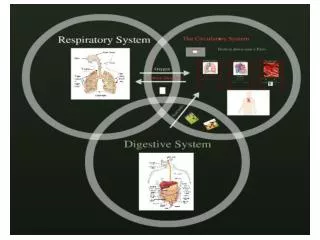
Mechanics of respiration. Dr.Kalpana. Specific Learning objectives. At the end of class students should be able to: Explain the muscles involved in breathing Explain pressure changes during ventilation Explain the role of lung surfactant. The Respiratory System. The Mechanics of Breathing.

E N D
Mechanics of respiration Dr.Kalpana
Specific Learning objectives • At the end of class students should be able to: • Explain the muscles involved in breathing • Explain pressure changes during ventilation • Explain the role of lung surfactant
The Respiratory System The Mechanics of Breathing Mechanics of Breathing
VENTILATION • Breathing in – inspiration / inhalation • Breathing out – expiration / exhalation
Ventilation • Occurs because the thoracic cavity changes volume • Insipiration uses external intercostals and diaphragm • Expiration is passive at rest, but uses internal intercostals and abdominals during severe respiratory load • Breathing rate is 10-20 breaths / minute at rest, 40 - 45 at maximum exercise in adults
MUSCLES OF INSPIRATION • Diaphragm - increases the vertical diameter of chest wall (75% of inspiration) • External intercostals - increase the transverse and A-P diameters of chest wall (25% of inspiration)
Diaphragm is the main muscle of inhalation • When the diaphragm contracts, it pulls from the ribcage, flattening out • Inhalation is also assisted by the externalintercostals muscles • Connect higher rib to lower • When the muscle contracts, it causes expansion in the ribcage in all directions
ACCESSORY MUSCLES OF INSPIRATION • Sternocleidomastoid • Scaleni • Pectoralis minor ( Work during forced inspiration, exercise, airway obstruction)
EXPIRATION • Passive process • Results from elastic recoil of chest wall and lungs
MUSCLES OF FORCED EXPIRATION • Abdominal recti, transversus abdominis, obliques. • Internal intercostals
Internal intercostal muscle Abdominals
Movement of the Diaphragm Figure 17-9b
Movement of the Rib Cage during Inspiration Figure 17-10a
Movement of the Rib Cage during Inspiration Figure 17-10b
Inspiration • Inspiration-Active process • Contraction of these muscles increase the size of the thoracic cavity . • Boththe pleura will expand accordingly. • Diaphragm & Intercostal muscles • Diaphragm contracts increased thoracic volume vertically. • External Intercostals contract, expanding rib cage • They elevate the ribs upward and outward through pump handle and bucket handle movement. Lateral and anteroposterior dimension of the thorax. • More volume -> lowered pressure -> air in. • (Negative pressure breathing.) • Intrapulmonary pressure decreases (758 mm Hg
Inspiration 1) Our diaphragm pulls down 4) Air is sucked through the tubes into the lungs 2) Our intercostal muscles contract 3) Our chest expands Mechanics of Breathing
Inspiration Figure 22.13.1
Expiration • Expiration • Due to recoil of elastic lungs. • Passive. • Less volume -> pressure within alveoli is above atmospheric pressure -> air leaves lungs. • Note: Residual volume of air is always left behind, so alveoli do not collapse.
Expiration Figure 22.13.2
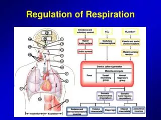
Pressures That Cause the Movement of Air In and Out of the Lungs(Pressure changes during ventilation)
Rib Cage Contract IntercostalsContractto Lift Spine Rib Volume Ribs Diaphragm Volume Mechanisms of Breathing: How do we change the volume of the rib cage ? • To Inhale is an ACTIVE process • Diaphragm • External Intercostal Muscles
Modes of Breathing • Quiet Breathing • Inhalation requires muscles • Contraction of diaphragm (75%), external intercostals (25%) • Exhalation passive • Lungs recoil due to elasticity • Forced Breathing • Inhalation • Accessory muscles include sternocleidomastoid and scalenes (muscles of the neck) • Exhalation • Internal intercostals, abdominal muscles
Pleural Cavity Very small space Maintained at negative pressure Transmits pressure changes Allows lung and ribs to slide Lungs Gas Exchange Chest Wall (muscle, ribs) Diaphragm (muscle) Pleural CavityImaginary Space between Lungs and chest wall
Pleural fluid produced by pleural membranes • Acts as lubricant • Helps hold parietal and visceral pleural membranes together
Pressure Relationships in the Thoracic Cavity • Respiratory pressure is always described relative to atmospheric pressure • Atmospheric pressure (Patm) - pressure exerted by the air surrounding the body • Intrapulmonary pressure (Ppul) – pressure within the alveoli • Intrapleural pressure (Pip) – pressure within the pleural cavity
Intra pulmonary pressure pressure changes in the alveoli normal value -2 to -3 mmHg to +2to +3 mmHg Intrapleural pressure pressure in the pleural space normal value -6 mmHg to -3 mmHg Significance –prevent collapse of lung and facilitates in venous return. Transmural pressure pressure difference across the lung
Intrapulmonary Pressure • Also called intra-alveolar pressure • Is relative to Patm • In relaxed breathing, the difference between Patm and intrapulmonary pressure is small: • about —2 mm Hg on inhalation or +2 mm Hg on expiration
The Respiratory Pump • Cyclical changes in intrapleural pressure operate the respiratory pump: • which aids in venous return to heart
REST PRESSURES Atmosphere Airways 760 MM Hg Intrapleural pressure Pleural Sac 756 mmHg Thoracic Wall Lungs
AIR IS A COMPRESSABLE GAS WHICH OBEYS BOYLE’S LAW • P1V1 = P2V2 • If Volume increases, Pressure must decrease • As lungs expand, pressure inside falls
INSPIRATION • Elevation of ribs expands lungs • Lowering of diaphragm by contraction also expands lungs • Expansion of lungs causes pressure inside to drop below atmospheric pressure • Air rushes in to fill the expanded lungs
INSPIRATION Atmosphere Airways 760 mm Hg 758mm Hg Intrapleural pressure Pleural Sac 754 mmHg Thoracic Wall Lungs
The lung is an elastic structure that collapses like a balloon and expels all its air through the trachea whenever there is no force to keep it inflated. • There are no attachments between the lung and the walls of the chest cage, except where it is suspended at its hilum from the mediastinum.
EXPIRATION • Return of ribs to rest position causes diminishing of lung volume • Return of diaphragm to rest position also causes diminishing of lung volume • Diminishing of lung volume causes pressure in lung to raise to a higher value than atmospheric pressure • Air flows out of the lungs
EXPIRATION Atmosphere 760 mm Hg Airways 762 mm Hg Intrapleural pressure Pleural Sac 756 mmHg Thoracic Wall Lungs
Lung "floats" in the thoracic cavity, surrounded by a thin layer of pleural fluid that lubricates movement of the lungs within the cavity. • There are two layers that contribute to the pleural cavity: • Parietal pleura • Visceral pleura.
Intrapulmonary and Intrapleural Pressures • During inspiration, intrapulmonary pressureis about -3 mm Hg pressure; during expiration is about +3 mm Hg • Positive transmural pressure(intrapulmonary minus intrapleural pressure) keeps lungs inflated
Pressure in the Pleural Cavity Pneumothorax results in collapsed lung that can not function normally Figure 17-12b
The pleural fluid (∼15–20 ml) forms a thin layer between the pleural membranes and prevents friction between surfaces during inspiration and expiration. • Continual suction of excess fluid into lymphatic channels maintains a slight suction between the visceral and the parietal pleural surface.
Pleural Pressure and Its Changes During Respiration • Pleural pressureis the pressure of the fluid in the thin space between the lung pleura and the chest wall pleura. • The normal pleural pressure at the beginning of inspiration is about -5 centimeters of water. • During normal inspiration, expansion of the chest cage creates more negative pressure -7.5 centimeters of water.
Injury to the Chest Wall • Pneumothorax: • allows air into pleural cavity • Atelectasis: • also called a collapsed lung • result of pneumothorax
Alveolar Pressure • Alveolar pressure is the pressure of the air inside the lung alveoli. • When the glottis is open and no air is flowing into or out of the lungs, the pressures in all parts of the respiratory tree, all the way to the alveoli, are equal to atmospheric pressure (0 cm water pressure).