Download
1 / 48
480 likes | 769 Views
Gastrointestinal Assessment. jeopardylabs.com/play/gi-jeopardy11. Anatomy . Abdomen. Anatomy and Physiology. Abdominal Landmarks: Abdominal wall is divided into four quadrants by a vertical and a horizontal line bisecting at the umbilicus. Nine Abdominal Regions. Match the Organs!.

E N D
Anatomy Abdomen
Anatomy and Physiology • Abdominal Landmarks: • Abdominal wall is divided into four quadrants by a vertical and a horizontal line bisecting at the umbilicus
Match the Organs! • Word Bank • Spleen • Stomach • Liver • Cecum • Gallbladder • Sigmoid Colon • Bladder • Descending colon • Ascending colon • Appendix • Small Intestine • Pubic Symphysis
Organs per Quadrant Right Upper Quadrant Left Upper Quadrant • Liver • Gallbladder • Duodenum • Head of pancreas • Right kidney and adrenal gland • Hepatic flexure of colon • Part of ascending and transverse colon • Stomach • Spleen • Left lobe of liver • Body of pancreas • Left kidney and adrenal gland • Splenic flexure of colon • Part of transverse and descending colon
Organs per Quadrant Right Lower Quadrant Left Lower Quadrant • Cecum • Appendix • Right ovary and tube • Right ureter • Right spermatic cord • Part of descending colon • Sigmoid colon • Left ovary and tube • Left ureter • Left spermatic cord
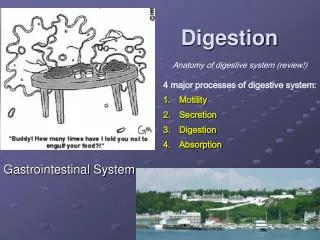
Assessment Gastrointestinal
Subjective Assessment • Changes in appetite? • Dysphagia? • Food intolerance? • Abdominal pain? • Nausea/vomiting? • Bowel Habits? • Diarrhea or constipation? • Changes in weight? • Past abdominal history? • Last bowel movement? • Nutritional assessment • Medication reconciliation
Objective Assessment • Examination Order: • Inspect • Auscultate • Percuss • Palpate • Why is the order of assessment techniques different?
Answer • During the physical examination of the abdomen, auscultation is performed prior to percussion or palpation because those techniques can increase peristalsis, providing a false interpretation of bowel sounds
Inspection • Position the patient supine with the head on a pillow • Ensure patient has emptied his or her bladder • Demeanor: • Benign facial expression • Slow, even respirations • Free from restlessness or absolute stillness
Inspection • Skin color, scars, or lesions • Should appear smooth, even color. • A common pigment variation is striae(stretch marks). Silvery, white jagged marks 1-6cm long. • Occurs following rapid or prolonged stretching, as in pregnancy or excessive weight gain. • Hair distribution • Pubic hair: Diamond shaped in adult males; inverted triangle in females
Inspection • Symmetry: Bilateral symmetry upon both inspiration and expiration, free from bulging or masses, which can be identified by shadows • Umbilicus: Midline, inverted without discoloration, inflammation, or hernia. • Contour: Examine the contour of the abdomen from the rib margin to the pubic bone. • Should range from flat to rounded
Inspection • Pulsations or Movement: • Aortic pulsation in the epigastric area • Particularly in thin people with good muscle wall relaxation • Respiratory movement • Particularly in males • Peristalsis • Slow waves ripple across the abdomen
Auscultation • Bowel sounds are caused by the movement of air and fluid through the small intestine • Begin listening at the: • RIGHT LOWER QUADRANT (RLQ)This is the ileocecal valve area and bowel sounds are always present here, normally.
Bowel Sounds Auscultation • Bowel sounds are high pitched, gurgling, with irregular occurrence between to 5-30 times a minute • Hyperactive: Greater than 30/minute • Normative: Between 5-30/minute • Hypoactive: Less than 5/min • Hyerperistalsis: (growling stomach), Borborygmus • To say no bowel sounds are present, one much auscultate for a FULL FIVE minutes. A perfectly “silent” abdomen is uncommon. http://evolvels.elsevier.com/section/default.asp?id=2259_global_0001&mode=
Auscultation of Vascular Sounds • Auscultate for vascular sounds or bruits, especially in the patient with hypertension • Use firmer pressure over the aorta, renal arteries, iliac, and femoral arteries • Generally NOT present http://evolvels.elsevier.com/section/default.asp?id=2259_global_0001&mode=
Percussion • Why? • Assess density of abdominal contents • Locate organs • Screen for abnormal fluid or masses • Expected findings: • Tympany: Over majority of the abdomen due to air rising to the surface when the patient is supine • Dullness: Occurs over organs (liver), distended bladder, adipose tissue, fluid, or a mass • Hyperresonance: Present with gaseous distention
Percussing the Liver • 1). Measure liver height: • Start at the right midclavicular line: • Percuss in the area of lung resonance • Percuss down the interspaces until the sound changes to a dull quality • Mark the spot of change • (generally the 5th intercostal space) • 2). Then, • Percuss abdominal tympany • Percuss UP along the midclavicular line • Mark where the sound changes from tympany to dull • 3). Finally, • Measure the distance between the two marks. Generally, the liver span ranges between 6-12cm, correlating with height. • Hepatomegaly: Enlargement of the liver
Fist Percussion: CVA Tenderness • CVA: Costovertebral Angle Tenderness • Patient should be sitting up (90’), stand behind to perform percussion. • Process: • Place one hand over the 12th rib at the costovertebral angle of the back. • Thump that hand with the ulnar edge of your other fist • Generally, patient will feel a thump, but no pain. • Sharp pain indicates inflammation of the kidney or pananephric area (near the kidney).
Fluid wave • Used when ascites is suspected • How: • Place patient’s hand firmly on the abdominal midline • Place left hand on patient’s right flank • Using right hand, give the left flank a firm strike • If ascites is present, a distinct tap will be felt on your left hand
Palpation • Why palpate? • Assess the size, location, and consistency of organs • Screen for abnormal masses or tenderness • Watch for: • Muscle guarding • Rigidity • Large masses • Tenderness
Palpation • Types of Palpation: • Light palpation: Keep four fingers together, depress skin lightly by approximately 1cm • Deep palpation:Press down 5-8cm. • Bimanual palpation:Using two hands, the bottom hand senses, while the top pushes. Used with the large or obese abdomen. • How: • Have the patient lay supine with knee’s slightly bent • Place the palpating hand low and parallel to the abdomen • Palpate to desired depth with four fingers together • Make a gentle, rotary motion • Move clockwise, lifting fingers completely off the skin • Save palpation of tender areas until last
Palpation: Abnormal Findings • If you identify a mass, distinguish from a normally palpable structure or enlarge organ: • Note: • Location • Size • Shape • Consistency (soft, firm, hard) • Surface (smooth, nodular) • Mobility (including with respirations) • Pulsatility • Tenderness
Aortic Pulsation • Using opposing thumb and fingers, palpate the aortic pulsation • Location: Upper abdomen, left of midline • Generally 2.5-4cm, anterior pulsation
Assessing Rebound Tenderness • Also known as Blumberg’s Sign • Used when patient: • reports abdominal pain • reports tenderness during palpation • How: • Perform at the end of assessment • Select a location away from reported pain • Hold hand 90’ to abdomen • Push into abdomen slowly and deeply • Lift hand up quickly • Response: • No pain on release of pressure: Expected • Pain: sign of peritoneal inflammation, possible appendicitis
Age Related Variations • Infant • Diastasis recti: Separation of rectus muscles with a visible bulge along the midline. Most common with black infants, disappears by early childhood • Child • Protuberant abdomen: When supine and standing, children under age 4; flat when supine after age 4 • Movement with respirations: Until age 7
Age Related Variations • Older adult • Subcutaneous fat on abdomen and hips • Liver is easier to palpate due to decreased abdominal muscle tone • Liver location is palpated 1-2cm below costal margin upon inhalation
Anus, Rectum & Prostate Anatomy & Physiology
Structure & Function • Anus: • The outlet of the gastrointestinal tract • 3.8cm long • Has two sphincters: • Internal: Involuntary • External: Voluntary • Rectum: • Distal portion of the large intestine • 12cm long; from sigmoid colon, 3rd sacral vertebra
Structure & Function • For Him: • Prostate: • Lies in front of the anterior wall of the rectum • Secrets thin, milky alkaline fluid that helps sperm viability • For Her: • Uterine Cervix: • Lies in front of the anterior wall, may be palpated through it
Anus, Rectum & Prostate The Assessment
Subjective • Usual bowel routine? • Change in bowel habits? • Rectal bleeding, blood in stool? • Self-care behaviors • Family history • Rectal conditions: • Itching • Hemorrhoids • Fissure • Fistula • Medications
Objective Assessment • Inspection: • Color: • Moist, more pigmented skin than perianal skin • Surface characteristics: • Coarse folded skin • Hair: • None • Hemorrhoids: • Flabby skin sac; shiny blue skin sac: thrombosed hemorrhoid • Dimpling, inflammation, swelling, hair tuft, or tenderness at the tip of coccyx may indicate pilonidal cyst (resulting from inflammation from ingrown hair, debris) • Lesions: • Abnormal finding with inflammation; document location using 12hour clock method.
Objective Assessment • Positioning: • Females: • Left lateral decubitus • Lithotomy (if examining genitalia too) • Males: • left lateral decubitus • standing
Objective Assessment Place the pad of index finger gently against anal verge Feel for the tightening of sphincter, then relaxation; as it relaxes, flex the tip of your finger and insert slowly in the direction of the umbilicus NEVER use 90’ angle! • Palpation: • Gloves! Lubricating jelly. • Explain what you are doing!
Objective Assessment • Rotate your examining finger, palpate entire muscular ring • *assess sphincter tone • Canal should feel smooth and even • Ask person to tighten evenly around finger • Bidigital palpation: Use thumb against the perianal tissue. Press examining finger; assess swelling or tenderness. • Also, assesses bulbourethral glands • *Cowper’s glands, in males only
Stool Assessment • Occult Blood • Test may vary by agency or institution. • Positive is an abnormal finding • False-positive may be caused after eating large amounts of red-meat in past 3 days
Age & Situational Variations • Newborns: • Visual inspection of anus • Confirm patent rectum with meconium passing • Meconium: greenish stool passed the first 24-48hrs after birth • Infants & Children: • Buttocks should be firm and rounded, no masses or lesions • Meningocele • Mongolian spots • Diaper rash • Omit palpation unless symptoms warrant • If necessary: child on back, legs flexed- use fifth finger due to size
Health Promotion • After age 50… • Colorectal screening: • Digital rectal exam annually • Fecal occult blood test annually • Sigmoidoscopy every 5 years • Colonscopy every 10 years • Prostate cancer screening: After age 45 in black males; after age 50 all others • Prostate-specific antigen (PSA) annually
quizzes http://evolvels.elsevier.com/section/default.asp?id=2259_global_0001&mode= http://evolvels.elsevier.com/section/default.asp?id=2259_global_0001&mode=