Download
1 / 43
440 likes | 646 Views
Nervous System 2 Cerebrovascular Disease. Prof John Simpson. Cerebrovascular disease (CVD). “strokes” brain disease due to vascular pathology thrombosis, embolism or hypotension causing ischaemia/hypoxia haemorrhage causing disruption

E N D
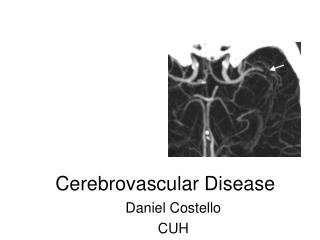
Nervous System 2Cerebrovascular Disease Prof John Simpson
Cerebrovascular disease (CVD) • “strokes” • brain disease due to vascular pathology • thrombosis, embolism or hypotension causing ischaemia/hypoxia • haemorrhage causing disruption • major cause of death and disability, especially in more developed countries • commonly associated with atheroma, diabetes and hypertension
Two major pathologies • infarction • thrombotic (overall 80%+ of all strokes) • embolic • hypotensive • (venous) • haemorrhage • intracerebral • subarachnoid • but, one can lead to the other!
Hypoxia and the brain • brain highly oxygen (and glucose) dependent • blood flow normally autoregulated • problems arise from 1) major fall in BP or systemic hypoxia causing diffuse damage or 2) vessel blockage, causing focal damage
Diffuse hypoxic damage • depends on severity and duration of hypoxia • most susceptible neurons in hippocampus, Purkinje cells, cerebral cortex • affected brain oedematous, raising ICP • causes anything from mild confusion to PVS to immediate brain death • in acute hypotension, may also be focal damage • “watershed” (border zone) infarcts – most often between anterior cerebral and middle cerebral artery supplies
Focal hypoxic damage • results depend on presence of collaterals • some exist on surface, e.g. Circle of Willis • but not within brain • focal vascular abnormality due to • thrombosis or embolism • clinical effects ~ site, extent and speed of onset of vascular block
Thrombotic causes of focal hypoxia • mostly atheroma - commoner in DM and HT • usually thrombosis at carotid bifurcation, origin of middle cerebral artery or in basilar artery • vasculitis • infective (more so in immunosuppressed • syphilis, TB, fungi, toxoplasmosis • autoimmune disease • hypercoagulable states • dissecting aortic aneurysms • drug abusers • trauma • cardiac or respiratory arrest
Embolic causes of focal hypoxia • commonest are cardiac mural thrombi • MI, valvular disease, atrial fibrillation • arterial thromboemboli - especially from carotid plaques (sometimes include plaque material) • paradoxical emboli - children with cardiac anomalies • emboli of other material (tumour, fat, marrow, air)
Cerebral embolism • middle cerebral territory most often affected • emboli lodge at branches or stenoses • often, occlusion cannot be identified PM • ?thromboemboli already lysed • “shower” embolism of fat may occur after fractures • capillary blockages – disturb higher cortical function and consciousness, often with no localizing signs • widespread haemorrhagic lesions of white matter characteristic of bone marrow embolism after trauma • tumour emboli more important as source for metastases, then cause of hypoxia
Cerebral infarcts • sometimes classified as red or pale • depends on presence of haemorrhage from infarcted vessels • (any infarct may show surrounding zone of lesser hypoxic damage and hyperaemic reaction, which may be oedematous) • venous infarcts – usually beside sinuses – associated with infection, dehydration and drugs (oral contraceptives)
Natural history of infarcts • effects depend on site, size and speed of onset • in some effect complete from the start, in others clinical picture evolves • thrombotic infarcts most commonly internal capsule (corticospinal paths), hence hemiplegias etc • reperfusion (micro)haemorrhages may occur • if patient survives, infarcted tissue phagocytosed by microglia and monocytes from blood, then gliosis • macrophages persist at site for years as lipid-containing “compound granular corpuscles” • in red infarct, macrophages also contain iron • end result of repair often a cystic cavity with gliotic wall
increased eosinophilia of neurons then neuronal death and cell infiltrate eventual gliosis Microscopic changes in infarct
Intracranial haemorrhage • secondary • following infarction • primary • extradural and subdural • usually traumatic in origin • subarachnoid and intraparenchymal (aka intracerebral) • usually due to vascular disease
Subarachnoid haemorrhage • most often due to cerebral artery berry (saccular) aneurysms • but also by extension from intracerebral haemorrhages or due to bleeding diseases, trauma, tumour, vasculitis etc
Berry (saccular) aneurysms • incidental finding in ~ 2% of post-mortem examinations, multiple in maybe a third • occur near major branch points on Circle of Willis or just beyond • more common on anterior part of Circle or its branches
Aetiology of berry aneurysms • genetic factors may be important in some cases • e.g. increased risk in ADPKD, Ehlers-Danlos syndrome, Marfan’s syndrome) etc • cigarette smoking and hypertension also predisposing factors • “congenital”, but not present at birth, though underlying defect in media may be
Berry aneurysms • thin-walled out-pouching • usually < 1 cm diameter • wall consists only of intima • rupture at apex, usually into subarachnoid space, but sometimes into brain or both
Berry aneurysms • rupture most often in 40- 50s • may be precipitated by sudden ICP rise • also by hypertension • typically sudden severe headache and rapid loss consciousness • ~ 10-15% die, but most recover consciousness in minutes • may show meningism • rebleeding common and makes prognosis worse
Subarachnoid haemorrhage • early effects include • increased risk of vasospasm of other vessels • can lead to additional ischemic injury, espec. if spasm involves Circle of Willis • presumably due to vascular mediator • late sequelae • meningeal fibrosis and scarring • possible obstruction of CSF flow/reabsorption.
CSF in subarachnoid haemorrhage • initially bright red blood • later, xanthochromia as red cells degenerate
Intraparenchymal (intracerebral or cerebral) haemorrhage • 80 % death rate • sudden onset, causing rapid rise in ICP • 50%+ associated with hypertension • ? microaneurysms (of Charcot-Bouchard) • ? just arteriosclerotic branch points • remainder due to vascular malformations, bleeding disease, vasculitis etc
Intracerebral haemorrhage • usually affects basal ganglia, brainstem, cerebellum or cerebral cortex • major tissue disruption and destruction • may extend into ventricles and/or subarachnoid space • in survivors, haematoma surrounded – like infarcts - by zone of reaction, then repair with gliosis
Other causes of haemorrhage angiomas, AV malformations etc
Hypertension and CVD • common cause of CVD • frequently associated with atheroma and diabetes • responsible for - • intracerebral haemorrhage • and rupture of berry aneurysms, so subarachnoid haemorrhage • lacunar infarcts • hypertensive encephalopathy • acute or chronic
Hypertension and lacunar infarcts • arteriosclerosis +/- occlusion of vessels supplying basal ganglia, hemispheres and brainstem • causes single/multiple small cavitated infarcts (“lacunes”) • tissue loss with scattered compound granular corpuscles surrounded by gliosis • clinical effects depend on location - may be “silent”
Acute hypertensive encephalopathy • syndrome of diffuse cerebral dysfunction • headaches, confusion, vomiting and convulsions, sometimes leading to coma • usually part of “malignant” phase hypertension • rapid treatment needed to reduce raised ICP • at PM, oedematous brain +/- tentorial or tonsillar herniation • arteriolar fibrinoid necrosis and petechiae throughout brain
Chronic hypertensive encephalopathy • one cause of vascular (multi-infarct) dementia • dementia often with focal neurological defects • caused by multifocal vascular disease over long time • cerebral atheroma • thrombosis or embolism from carotids or heart • cerebral hypertensive arteriolosclerosis
Intracranial vascular pathology in summary • Extradural and subdural haemorrhage • trauma • Subarachnoid haemorrhage • berry aneurysms • Intracerebral haemorrhage • hypertension • Cerebral infarction • atheroma/thrombosis/embolism